Access a toolkit of functional, consistent in vitro models to study neurodegenerative disease
Powered by opti-ox™
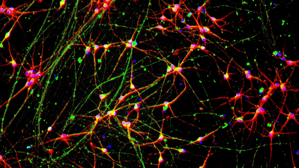
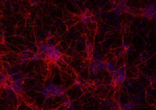
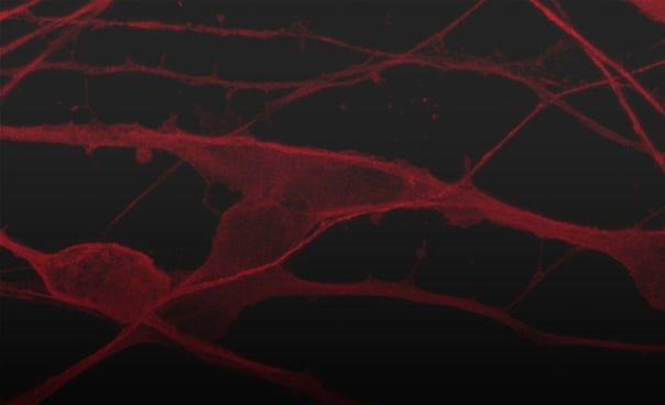
ioGlutamatergic Neurons rapidly acquire a homogeneous neuronal phenotype upon thawing, as captured in this 7-day time course. Powered by opti-ox technology, these cells consistently convert to functional excitatory neurons characterised by >80% expression of glutamate transporter genes VGLUT1 and VGLUT2.
Delivered cryopreserved and ready-to-culture, ioGlutamatergic Neurons offer a highly defined, easy-to-use human model for translational research and drug discovery.

Bulk RNA-sequencing analysis of three independent lots of ioGlutamatergic Neurons reveals tight clustering at specific timepoints, demonstrating the manufacturing precision of opti-ox technology. Analysis of differentially expressed genes (|logFC| >0.5 and FDR <0.01) confirms no statistically significant variance between lots, ensuring users can rely on uniform performance and reproducible experimental data across every vial.
Prof. Marius Wernig and Dr. Mark Kotter discuss the paradigm shift of transcription factor-mediated cell programming and how opti-ox technology is industrialising the process. The speakers outline how this approach overcomes traditional variability to enable the scalable, precise manufacturing of human cells.

Raster plots generated using the MaxTwo HD-MEA System capture the rapid development of functional networks in ioGlutamatergic Neurons co-cultured with human iPSC-derived astrocytes. Spontaneous activity observed at day 7 evolves into clear synchronised bursting by day 31, represented by vertical blue lines across 1,024 active electrodes. This confirms a rapidly maturing functional system ideal for assessing network dynamics and compound effects.

Co-culturing ioGlutamatergic Neurons, ioGABAergic Neurons, and hiPSC-derived astrocytes provides a physiologically relevant platform to study network hyperexcitability. As shown in the graphs (A) and raster plots (B), ioGABAergic Neurons functionally integrate to inhibit excitatory activity, reducing the number of spikes per network burst in a cell number-dependent manner. This robust multi-cell-culture can be used as a platform to model neurological conditions such as epilepsy, autism and schizophrenia in vitro.

ioGlutamatergic Neurons engineered with the PSEN1 M146L, APP V717I (London) and APP KM670/671NL (Swedish) mutations recapitulate the changes in Aβ peptide ratios observed in Alzheimer’s disease patients. This demonstrates their validity as an in vitro model to study Alzheimer's disease and for the discovery of drugs targeting the pathogenic Aβ pathway.

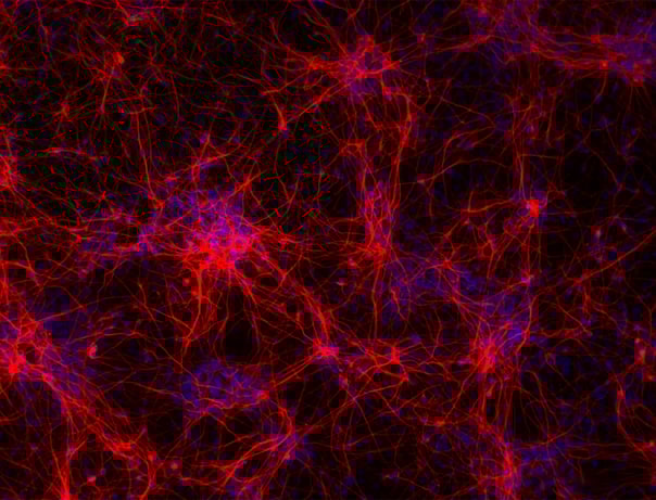
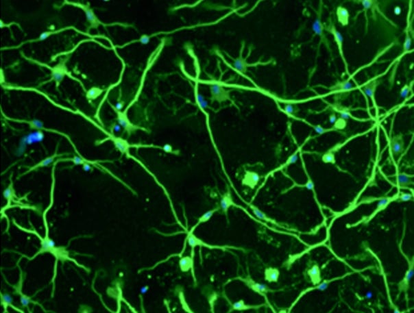
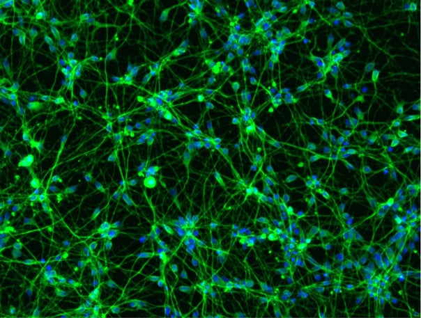
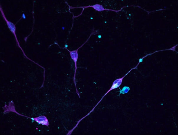
Charles River Laboratories leveraged the MaxTwo HD-MEA system to characterise bit.bio’s Huntington’s disease (HD) model. By comparing ioGlutamatergic Neurons HTT 50CAG/WT to their genetically-matched wild-type control, the team identified distinct functional phenotypes, including delayed network formation, decreased axonal branching, and reduced spontaneous activity. These results demonstrate the model's ability to recapitulate complex disease-related phenotypes, offering a valuable tool for screening.

ioGlutamatergic Neurons demonstrate high suitability for antisense oligonucleotide (ASO) efficacy screening following delivery by gymnosis. The lack of marked intra- or inter-plate variability confirms these cells as a robust, physiologically relevant model for validating therapeutic candidates.

ioGlutamatergic Neurons have been engineered to constitutively express green fluorescent protein (GFP), offering scientists an in vitro, fluorescent human neuronal model ideal for culturing with other cell types, enabling effortless tracking in multi-cellular systems.
Stable GFP expression enables easy, real-time tracking in complex, multi-cellular systems, facilitating the study of glial interactions or network function alongside inhibitory neurons. This makes the cells ideal for live-cell imaging to assess neurite outgrowth, morphology, and survival in response to compound treatment.

CRISPRko-Ready ioGlutamatergic Neurons reveal distinct phenotypic clustering in a pooled knockout screen targeting 100 neurodegenerative disease-relevant genes. As visualised in the UMAP, single-cell analysis shows that aminoacyl-tRNA synthetase (aaRS) knockouts—including AARS1 and GARS1—group together, while non-targeting controls remain evenly distributed. Pathway analysis indicates that targeting these genes activates the unfolded protein response (UPR), validating the model's ability to recapitulate mechanisms found in neurodegenerative conditions like Charcot–Marie–Tooth neuropathy (CMT).
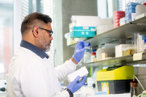
In this video, our scientist takes you through the step-by-step process of how to thaw, seed and culture ioGlutamatergic Neurons, which complements our expert scientist's top tips on understanding the importance of handling cells gently, preparation of coatings, media changes and cell density.

Dr Shushant Jain
Group Leader | In Vitro Biology | Charles River, 2021

Dr Mariangela Iovino
Senior Group Leader | Biology Discovery | Charles River

Dr Koby Baranes
Research Associate | University of Cambridge

Dr Jeremy Anton
Scientist | Charles River

Professor Deepak Srivastava
Professor | Molecular Neuroscience | King’s College London and Group Leader | MRC Centre for Developmental Disorders
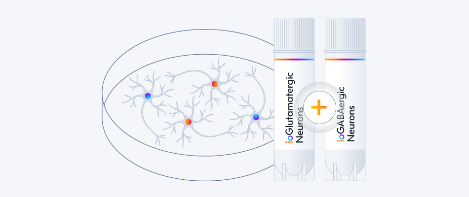
Study network synchrony, functional connectivity, and drug responses using MEA in a more physiologically relevant neuronal network with balanced excitatory and inhibitory activity.
Explore ioGABAergic Neurons
Explore ioGABAergic Neuron Disease Models
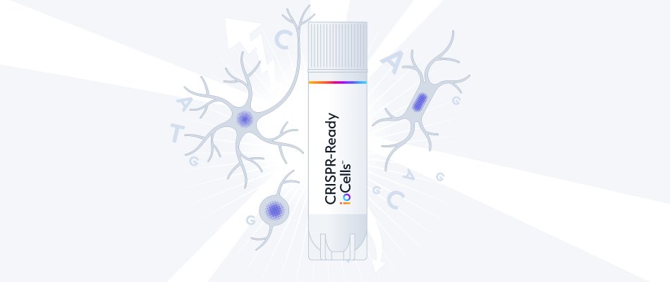
CRISPR-Ready ioCells stably express Cas9 nuclease or dCas9 variants and come with optimised cell culturing and guide delivery protocols.
Start measuring readouts from gene perturbations and CRISPR screens within days
Explore CRISPR-Ready ioCells
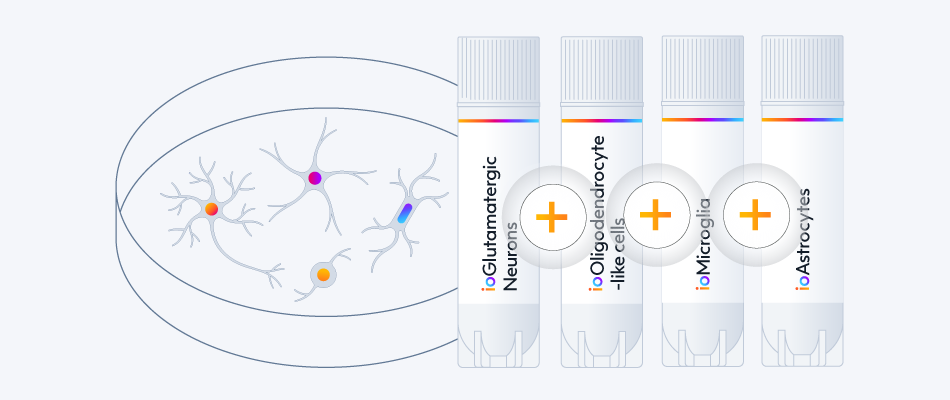
Access our in vitro neuroscience toolkit to develop complex models for investigating cellular communication and disease mechanisms in the human context.
ioMicroglia
ioOligodendrocyte-like cells
ioAstrocytes
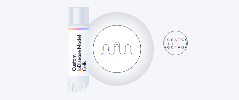
Study the impact of mutations related to neurodegenerative diseases in consistent, defined and scalable human CNS disease model cells with genetically matched controls.
- Alzheimer's disease
- Parkinson's disease
- Huntington's disease
- ALS and FTD


_MAP2(R)_DAPI(B)_%20(1).png?width=604&name=a-HTT50CAGWT_Overlay__TUBB3(G)_MAP2(R)_DAPI(B)_%20(1).png)


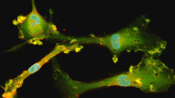
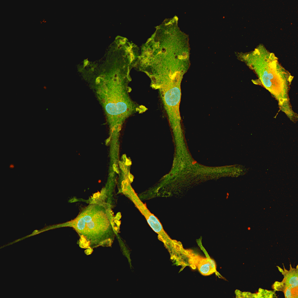

Hoescht(blue)TUBB3(blue)_day4.png?width=604&name=bit.bio_ioGlutamatergic%20Neurons_60xMAP2(red)Hoescht(blue)TUBB3(blue)_day4.png)


.png?width=604&name=a.HTT50CAGWT__TUBB3(G).png)


.jpeg?width=604&name=New%20Project%20(13).jpeg)


-1.png?width=604&name=CRL%20video%20%231%20card%20for%20webpage%20(thinner%20gradient%20line)-1.png)
Hoescht(blue)TUBB3(blue)_day4.jpg?width=604&name=bit.bio_ioGlutamatergic%20Neurons_60xMAP2(red)Hoescht(blue)TUBB3(blue)_day4.jpg)