Defined. Functional. Human.
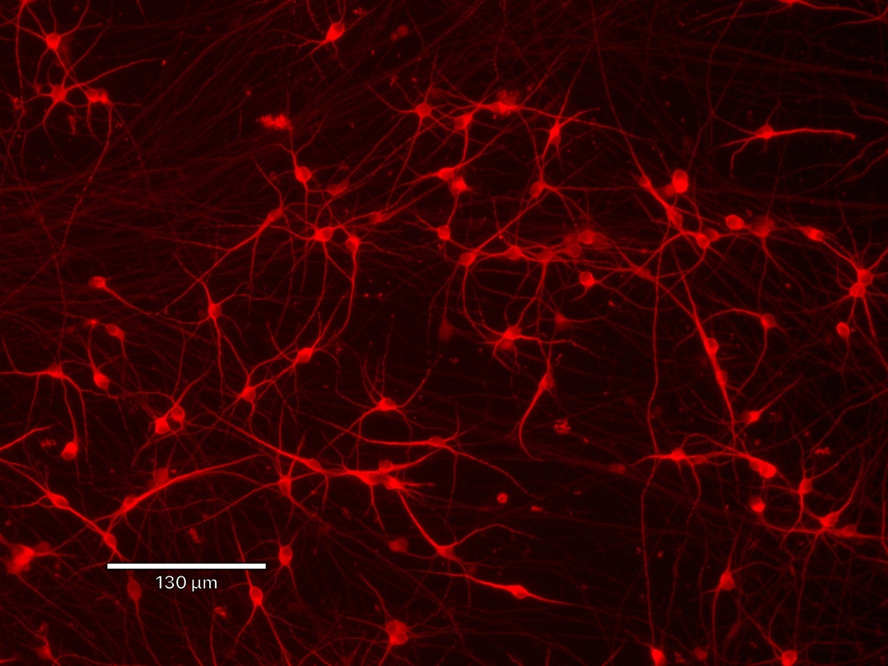
The predictive value of in vitro electrophysiology depends on the cell model. Variability across batches, between donors, and within cell populations in traditional animal-derived or iPSC-derived cells often introduces confounding biological noise, complicating the interpretation of results. ioCells, powered by opti-ox™ technology, provide consistent, defined populations of either human neurons or glia within days, giving researchers greater control over their experiments and reducing variability at the source.
Proven performance
Compatible with MEA assays (Axion, MaxWell Biosystems) and patch-clamp workflows across in vitro electrophysiology.
Defined inputs
Using consistent human neurons or glia reduces variable inputs from animal cells or mixed iPSC-derived cultures.
Experimental control
Defined cell identities and purity provide greater control over experimental design and interpretation.
ioCells in action
ioCells in action
Tracking functional neuronal maturation over time with HD-MEA
Maturation of neuronal network activity in ioGlutamatergic Neurons. The network activity of ioGlutamatergic Neurons was characterised using the MaxTwo HD-MEA System. The Axon Tracking Assay reveals the emergence of complex neural circuits through reconstructed axonal paths of travelling action potentials (left). Over the culture period, an increase in both total axon length (middle) and firing rate (right) was observed. Together, these data show the functional maturation of neuronal networks in physiologically relevant co-culture with human iPSC-derived astrocytes.
Data courtesy of Charles River Laboratories and MaxWell Biosystems.
Robust ion channel and receptor activity

Electrophysiological characterisation of ioSensory Neurons using medium throughput automated patch clamp analysis. Representative recordings from ioSensory Neurons show voltage-gated sodium (Nav) current-voltage relationships under (A) control and (B) tetrodotoxin (TTX) conditions, and (C) TTX concentration–response. (D) Example action potential firing measured in current clamp mode. Data from the “Characterising ioSensory Neurons on the Patchliner, an automated patch clamp system” application note from Nanion Technologies.
On the Patchliner, ioSensory Neurons achieved 100% capture and >80% seal rates. Recordings confirmed NaV currents (TTX-s and TTX-r), ligand-gated responses to GABA, glutamate, and P2X, heat-activated currents, and action potential firing under current clamp.
Defined neuronal model for studying neurological diseases

Functional excitatory–inhibitory network interactions in a human neuronal tri-culture model. ioGABAergic Neurons form functional networks with excitatory ioGlutamatergic Neurons and inhibit their activity in a cell number dependent manner (A). MEA analysis shows that ioGABAergic Neurons inhibit the excitatory activity of ioGlutamatergic Neurons in a ratio dependent manner (B), and that the excitatory–inhibitory balance can be further modulated by drugs targeting GABergic signalling.
ioGlutamatergic Neurons, ioGABAergic Neurons, and hiPSC-derived astrocytes provide a robust in vitro model to study network hyperexcitability and support the discovery of new therapies for neurological diseases, such as epilepsy, autism and schizophrenia.
Longitudinal HD-MEA analysis of ioMotor Neurons & astrocytes co-culture

Functional network maturation of ioMotor Neurons in co-culture with rat astrocytes. Representative multielectrode array (MEA) data shows neuronal activity in ioMotor Neurons co-cultured with rat cortical astrocytes over a 42 day period. (A) Percent active area: Quantitative analysis demonstrating a progressive increase in the percentage of the array area showing neuronal activity from Day 7 through Day 42. (B) Action potential tracking for individual neurons: Clump-free morphology enables single cell assessments of ioMotor Neurons. (C) Mean firing rate heatmaps: Comparison of spontaneous activity between Day 21 (left) and Day 42 (right). The heat maps show a significant increase in mean firing rates (Hz) over time, which is consistent with the progressive maturation of the functional motor neuron network.
This protocol details how to culture ioMotor Neurons with rat cortical astrocytes for MEA assays. It includes plate preparation, media recipes, and step-by-step instructions to support stable network activity and extended recordings of motor neuron function.
What scientists say about ioCells

Dr Elizabeth Di Lullo
Associate Scientific Director | Brainever
“We thawed the cells and they looked wonderful. Close to 100% viable and looked great in cell culture! No clumping and easy to culture.”
Recommended resources
_MAP2(R)_DAPI(B)_%20(1).png)
Ready to browse?
Explore our full portfolio of human iPSC-derived ioCells. From neurons and glia to muscle and immune cell types, our products are consistent and defined, supporting reliable data generation for applications in disease research and drug discovery.