Access a toolkit of human skeletal muscle cells to study neuromuscular disorders
Powered by opti-ox™
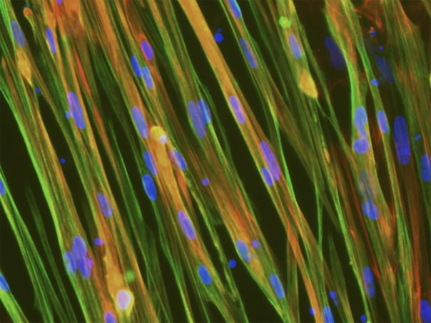
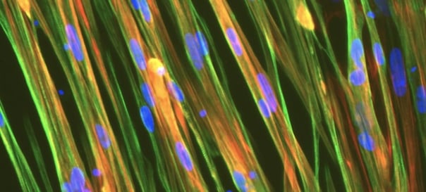
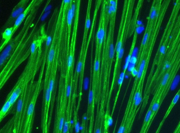
ioSkeletal Myocytes rapidly acquire a homogeneous phenotype upon thawing, as captured in this 10-day time course. Powered by opti-ox technology, these cells consistently convert into elongated, multinucleated human skeletal muscle cells expressing key myofilament proteins such as desmin and myosin heavy chain. Delivered cryopreserved, they provide a highly defined human model that is ready for physiological assays in just days.
Calcium imaging at day 10 post-thaw reveals the functional connectivity of ioSkeletal Myocytes. Following Fluo4-AM loading, the cells display robust spontaneous activity, with signal intensity reflecting intracellular Ca2+ flux. The video highlights the synchronised propagation of calcium waves across the monolayer, demonstrating the formation of a mature, physiologically relevant contractile network in a standard 2D format.

Cultured on the MUSbit platform (Bi/ond), ioSkeletal Myocytes self-organise into anchored 3D muscle bundles, as visualised by SEM at day 14 (A). Immunostaining reveals the progressive development of structural maturity over the culture period. By day 14, cells express key contractile markers, with high-magnification imaging displaying clear sarcomeric alpha-actinin cross-striations (B, yellow arrows), confirming the formation of highly organised muscle fibers.

In a functional validation by Bi/ond Solutions, wild-type and DMD exon deletion ioSkeletal Myocytes were cultured as 3D microtissues on the MUSbit platform. Compared to isogenic controls at day 14, the disease models (exon 44 deletion and exon 52 deletion) exhibit distinct functional deficits. Quantification reveals weaker contraction upon twitch and tetanus stimuli (A) and increased fatigue under sustained stimulation (B), confirming the 3D model's ability to recapitulate dystrophin-related pathology.

ioSkeletal Myocytes DMD Exon 44 Deletion cells demonstrate robust responsiveness to exon-skipping therapy following ASO delivery by gymnosis. ddPCR analysis (A) confirms a concentration-dependent increase in the in-frame skipped transcript (blue) and a corresponding decrease in the non-skipped transcript (yellow). This transcriptional modification translates to protein rescue, with high-content imaging (B) revealing a dose-dependent restoration of dystrophin expression compared to wild-type controls. These data validate the model’s utility for screening genetic therapies.

High-resolution confocal imaging validates the protocol for co-culturing ioSkeletal Myocytes with ioMotor Neurons. By day 30, the system displays distinct structural organisation, with MAP2-positive neurons (red) in the culture alongside Desmin-positive myocytes (cyan). The presence of acetylcholine receptors (yellow, alpha-bungarotoxin) confirms that this co-culture method supports the development of key neuromuscular features suitable for complex assay development.
In this video, our scientist takes you through the step-by-step process of how to thaw, seed and culture ioSkeletal Myocytes.

Dr Shushant Jain
Group Leader | In Vitro Biology | Charles River, 2021

Amy Rochford
PhD Neural Engineering and Bioelectronics | Cambridge University

Dr Michael Duchen
Professor of Physiology | University College London
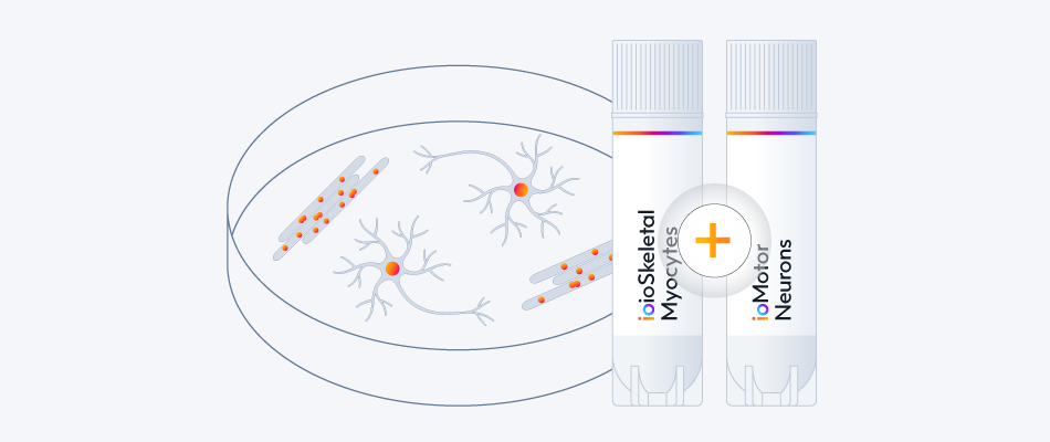
Study neuromuscular interactions and the impact of ALS-disease-related mutations by co-culturing skeletal myocytes and motor neurons. Access over 14 disease models and the single co-culture protocol.
View the co-culture protocol
Explore ALS & FTD Disease Models
Explore ioMotor Neurons
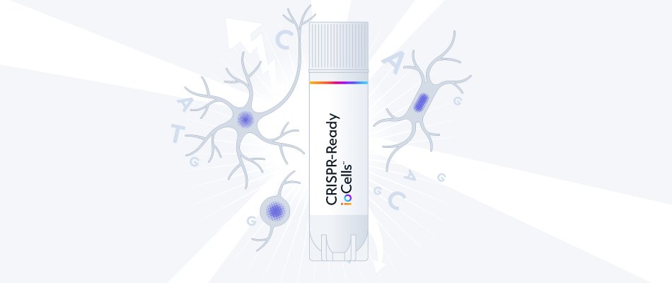
Interested in gene knockouts and CRISPR screens?
ioSkeletal Myocytes can be engineered to constitutively express Cas9 nuclease for the quick and easy generation of gene knockouts and CRISPR screens.
Learn about CRISPR screening services
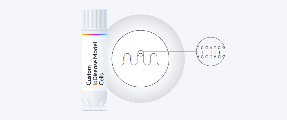
Build your custom disease model or reporter line to pair with wild-type ioSkeletal Myocytes as the genetically matched control.
Throughout the custom process, our experts will bring your project to life, and be on hand to support you with any technical queries.
Start the conversation today
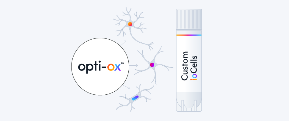
Interested in a new cell type?
Using opti-ox, bit.bio's scientists program iPSCs into defined identities. The result is a highly characterised, consistent model that offers reliability for research and drug discovery.


.png?width=604&name=ISSCR24-DMD-Exon44-3D-muscle-bundle%20(1).png)


.jpeg?width=604&name=New%20Project%20(13).jpeg)


/ioSkeletal-Myocytes-DMD-Exon-52-Deletion-hero.webp?width=1276&height=1190&name=ioSkeletal-Myocytes-DMD-Exon-52-Deletion-hero.webp)