Access a toolkit of functional, consistent in vitro models to study neurodegenerative disease and neuroinflammation
Powered by opti-ox™

Matteo Zanella, PhD
Associate Research Leader | Charles River
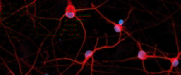
ioMicroglia rapidly acquire a homogeneous microglia phenotype upon thawing, as captured in this 10-day time course. This consistent and rapid maturation provides scientists with confidence in the reproducible generation of functional cultures in just a few days.
The HMC3 immortalised cell line has long been a widely used microglial model due to its ease of culture and scalability. However, it has some limitations that compromise its utility for translational research. HMC3 cells lack key microglial identity markers like P2RY12 and TREM2, possess a transcriptomic profile significantly different from primary microglia, and functionally demonstrate very weak or no phagocytic activity and a blunted cytokine response. In contrast, ioMicroglia provide a consistent, phenotypically accurate model, expressing key canonical markers and demonstrating robust phagocytic function and cytokine release profiles. Incorporating this human-relevant model offers a powerful opportunity to de-risk the drug discovery pipeline, filter out false positives, and increase the translational potential of preclinical findings.
ioMicroglia closely mimic the molecular identity of human foetal and adult microglia. They overcome the key limitations of sourcing constraints and donor variability of primary cells as well as the heterogeneity and batch-to-batch inconsistency caused by complex differentiation protocols.
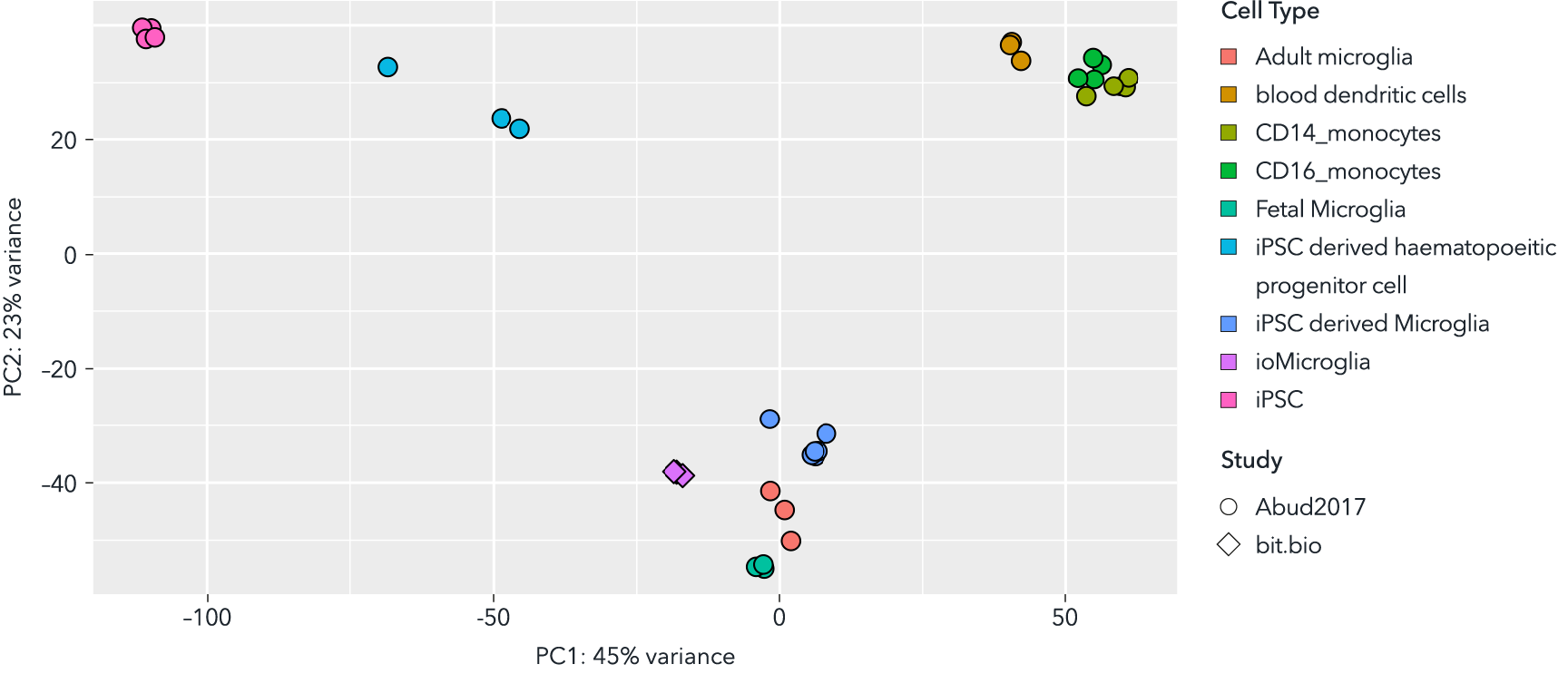
This principal component analysis (PCA) plot of bulk RNA sequencing data validates the transcriptomic identity of ioMicroglia. By integrating male donor-derived ioMicroglia with external data sets from Abud et al1., the analysis reveals that ioMicroglia exhibit a transcriptomic clustering with primary foetal and adult microglia. This demonstrates that ioMicroglia possess a molecular profile highly similar with in vivo human cells, providing an accessible, physiologically relevant model.

The cytokine release assay is a valuable tool for ensuring that microglia behave in vitro as they would in vivo. ioMicroglia secrete proinflammatory and anti-inflammatory cytokines (IL-6, TNFα, IL-1β,and IL-8) in response to stimuli such as Lipopolysaccharides (LPS) and Interferon Gamma (IFN-γ), and amyloidβ-42 (Aβ42).
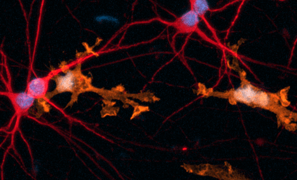
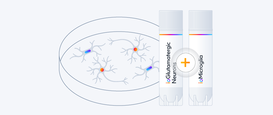
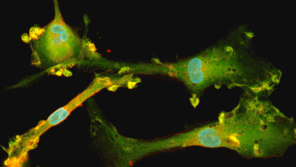
ioMicroglia demonstrate robust and selective phagocytic activity while in a 10-day co-culture with ioGlutamatergic Neurons. As captured in this time-lapse, the microglia actively engulf pHrodo Red Zymosan particles without any adverse effects on neuronal morphology, giving scientists confidence in their ability to build stable, functionally validated co-culture models for studying specific glial-neuronal interactions.
bit.bio has optimised protocols for phagocytosis and cytokine release assays, enabling scientists to implement co-culture systems and perform these assays with ease.
.png?width=600&height=329&name=AB42%20Percentage%20positive-%20Male%20WT%20ApoE%20(1).png)
ioMicroglia have been engineered with early-onset Alzheimer’s disease mutations in APOE and TREM2 genes, providing a system to study disease-related phenotypes. These models demonstrate robust phagocytic activity against both E. coli and fluorescent Aβ42 particles. Furthermore, the cells exhibit a complex cytokine secretion profile (including IL-6, IL-8, and IL-1β) in response to inflammatory stimuli (LPS/IFN-γ) and Aβ42 oligomers. This consistent, human-relevant model allows scientists to study how risk genotypes modulate core microglial functions.

A quad-culture model integrates ioMicroglia, ioOligodendrocyte-like cells, ioGlutamatergic Neurons, and human iPSC-derived astrocytes, providing a physiological system to investigate the role of glial cells in neurodegenerative disease mechanisms.

Human brain organoids often lack a functional immune component, limiting their ability to capture neuroimmune interactions. This application note demonstrates how integrating ioMicroglia enables a more physiologically relevant human 3D model with measurable inflammatory signalling, cytokine release, and pharmacological modulation.

ioMicroglia have been engineered to constitutively express green fluorescent protein (GFP), offering scientists a fluorescent human microglia model ideal for co-culture with other neural cell types, enabling effortless tracking in multi-cellular systems.
Constitutive GFP expression throughout the cytosol facilitates live-cell imaging, enabling the assessment of cell motility and visualisation of microglial activation states. GFP-labelled human microglia streamline cell sorting workflows by eliminating the need for antibody staining.

CRISPRko-Ready ioMicroglia enable high-resolution pooled single-cell CRISPR knockout (scCRISPR) screens to systematically map gene function. In a targeted study focused on 110 neurodegeneration-linked genes, cells were transduced and subsequently challenged with LPS to induce a transcriptomic activation signature.
In this video, our scientist takes you through the step-by-step process of how to thaw, seed and culture ioMicroglia, which complements our expert scientist's top tips on understanding the dynamic morphology of microglia, handling cells gently, and using the right seeding density and media changes.
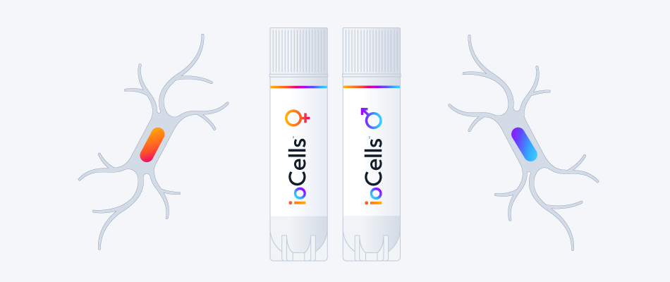
Women are greatly underrepresented in drug development and clinical trials.
Introducing female-derived cells into the early stage of research and drug discovery can help to better address this disparity.
Key applications for Female ioMicroglia in neurodegeneration drug discovery
- Neuroinflammatory in vitro modelling
- Target ID and validation
- Compound screening
Discover the data
Access 20 neuronal disease models and 4 microglia disease models with a single co-culture protocol.
View the co-culture protocol
Explore ioGlutamatergic Neuron Disease Models
Explore ioMicroglia Disease Models
Human iPSC-derived microglia engineered to constitutively express GFP enable easy visualisation, tracking and isolation of cells in complex multi-cell cultures.
Discover the data
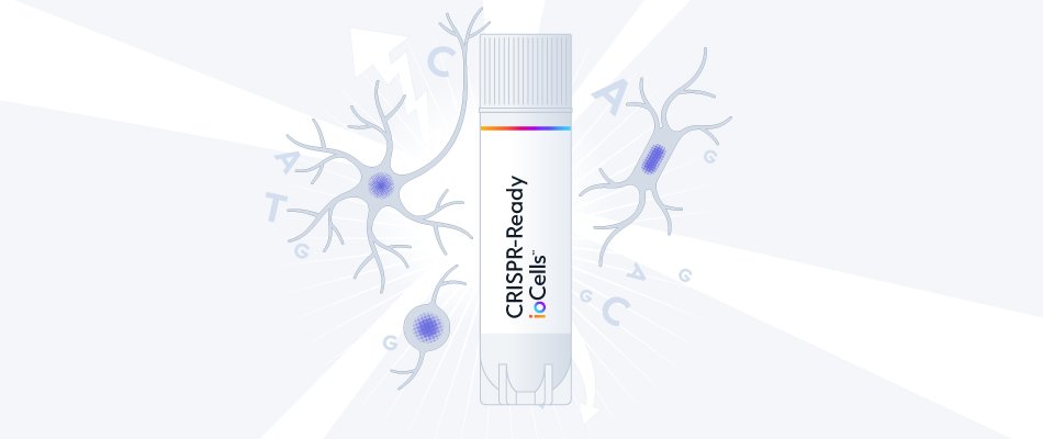
Built from our ioMicroglia Male and engineered to constitutively express Cas9.
With optimised guide RNA delivery protocols and high knockout efficiency, start measuring readouts from gene knockouts and CRISPR screens within days.
Save months of work by skipping complex cell line engineering and cell differentiation workflows.
Discover the data on CRISPRko-Ready ioMicroglia


Hoescht(blue)TUBB3(blue)_day4.png?width=604&name=bit.bio_ioGlutamatergic%20Neurons_60xMAP2(red)Hoescht(blue)TUBB3(blue)_day4.png)


_MAP2(R)_Tubb3(B)_Hoechst(B)_20x_merge-comp.jpg?width=604&name=Colour%20webinar%20with%20it-bio%20ioGlutamatergic%20Neurons_VGLUT2(G)_MAP2(R)_Tubb3(B)_Hoechst(B)_20x_merge-comp.jpg)