cat no | io1107 Early Access
Human iPSC-derived glutamatergic neurons constitutively expressing GFP
GFP ioGlutamatergic Neurons express key pan-neuronal and glutamatergic-specific markers
Immunofluorescent staining 11 days post-revival shows similar homogenous expression of pan-neuronal markers MAP2 and TUBB3 (upper panel) and the glutamatergic transporter VGLUT2 (lower panel) in both GFP ioGlutamatergic Neurons and the wild-type control.
The GFP signal is visible exclusively in GFP ioGlutamatergic Neurons and absent in the wild-type control (lower panel). 10X magnification.
Flow cytometry analysis of GFP expression in GFP ioGlutamatergic Neurons at day 11 and day 21
Flow cytometry analysis demonstrating GFP expression in >99% of cells for GFP ioGlutamatergic Neurons cultured until day 11 (centre), and no GFP expression seen in wild-type ioGlutamatergic Neurons (left). At day 21, the percentage of cells expressing GFP has not decreased, indicating there is no silencing of the reporter gene.
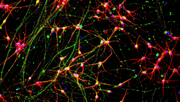
GFP ioGlutamatergic Neurons form structural neuronal networks by day 11
GFP ioGlutamatergic Neurons mature rapidly, show glutamatergic neuron morphology and form structural neuronal networks over 11 days, comparable to the wild-type control. Day 1 to 11 post thaw; 10X magnification.
GFP ioGlutamatergic Neurons demonstrate gene expression of neuronal-specific and glutamatergic-specific markers following deterministic programming
Gene expression analysis demonstrates that GFP ioGlutamatergic Neurons (GFP) and wild-type ioGlutamatergic Neurons (WT) lack the expression of pluripotency marker OCT4 (POU5F1) at day 11, while robustly expressing pan-neuronal (TUBB3 and SYP) and glutamatergic-specific (VGLUT1 and VGLUT2) markers, as well as the glutamate receptor GRIA4.
Gene expression levels were assessed by RT-qPCR, data normalised to HMBS; cDNA samples of the parental human iPSC line (iPSC) were included as reference. Data represents day 11 post-revival samples.
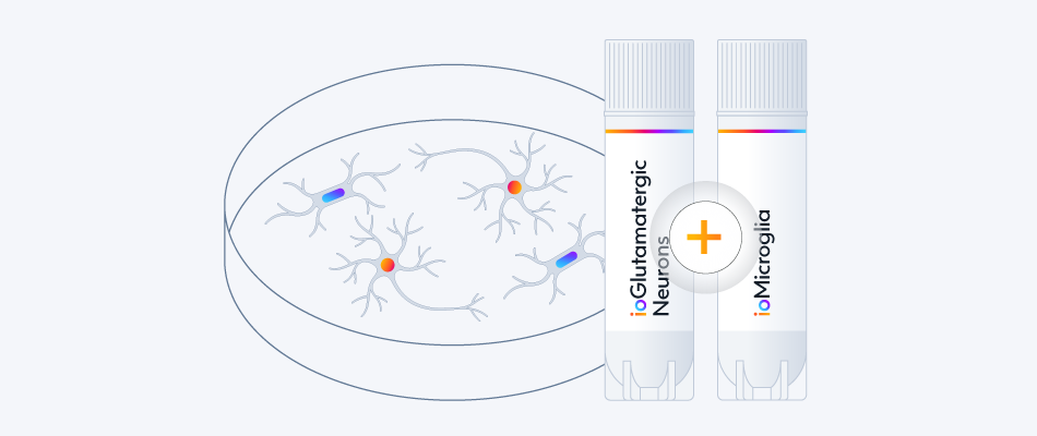
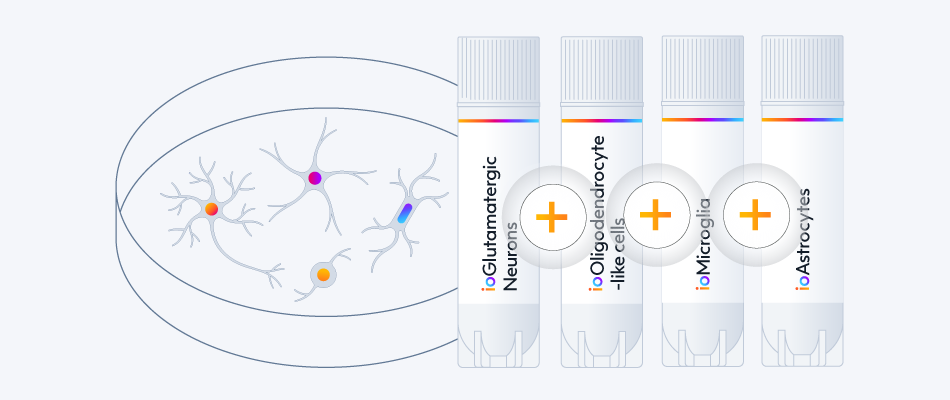
Easy-to-use co-culture protocol for GFP ioGlutamatergic Neurons with ioMicroglia
A maximum number of 20 vials applies. If you would like to order more than 20 vials, please contact us at orders@bit.bio.
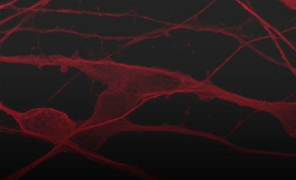
GFP ioGlutamatergic Neurons are a fluorescent human excitatory neuronal model derived from our well-established wild-type ioGlutamatergic Neurons. These cells are engineered to constitutively express green fluorescent protein (GFP), and provide a powerful, ready-to-use tool for diverse applications.
Stable GFP expression enables easy, real-time tracking in complex, multi-cellular systems, facilitating the study of cellular interactions with glia, or network function when co-cultured with inhibitory neurons. The cells are ideal for live cell imaging for assessment of neurite outgrowth, neuronal morphology and survival in response to compound treatment.
GFP ioGlutamatergic Neurons are delivered cryopreserved and ready-to-culture. This eliminates the time and effort required to engineer your own GFP-expressing iPSC lines and manage complex directed differentiation protocols.
Live-cell imaging ready
Assess neurite outgrowth, neuronal morphology and survival in real-time.
Co-culture compatible
Easily track GFP glutamatergic neurons in complex culture with glia.
Easy-to-use
Cells arrive programmed to mature rapidly upon revival. One medium is required in a two-step protocol.

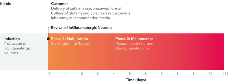
GFP ioGlutamatergic Neurons are delivered in a cryopreserved format and are programmed to mature rapidly upon revival in the recommended media. The protocol for the generation of these cells is a two-phase process: 1. Stabilisation for 4 days 2. Maintenance during which the neurons mature
Starting material
Human iPSC line
Karyotype
Normal (46, XY)
Seeding compatibility
6, 12, 24, 48, 96 & 384 well plates
Shipping info
Dry ice
Donor
Caucasian adult male, age 55-60 years old (skin fibroblast),
Genotype APOE 3/4
Vial size
Small: >1 x 10⁶ viable cells,
Evaluation pack*: 3 small vials of >1 x 10⁶ viable cells
Quality control
Sterility, protein expression (ICC), gene expression (RT-qPCR) and GFP expression (flow cytometry)
Differentiation method
opti-ox deterministic cell programming
Recommended minimum seeding density
30,000 cells/cm²
User storage
LN2 or -150°C
Format
Cryopreserved cells
Product use
ioCells are for research use only
Applications
Live-cell imaging
Co-culture
High-content screening
Neurotoxicology
Drug discovery
* Evaluation packs are intended for first-time users, or for existing users testing a new cell type or derivative. A user can request multiple evaluation packs as long as each one is for a different product, with only one pack allowed per product.
Dr Shushant Jain
Group Leader | In Vitro Biology | Charles River, 2021
Dr Mariangela Iovino
Senior Group Leader | Biology Discovery | Charles River
Dr Koby Baranes
Research Associate | University of Cambridge
Dr Jeremy Anton
Scientist | Charles River
Flow cytometry analysis of GFP expression at day 11 and day 21
Flow cytometry analysis demonstrating GFP expression in >99% of cells for GFP ioGlutamatergic Neurons cultured until day 11 (centre), and no GFP expression seen in wild-type ioGlutamatergic Neurons (left). At day 21, the percentage of cells expressing GFP has not decreased, indicating there is no silencing of the reporter gene (right).
GFP ioGlutamatergic Neurons exhibit comparable expression of neuron-specific markers to the wild-type control
Immunofluorescent staining 11 days post-revival shows similar homogenous expression of pan-neuronal markers MAP2 and TUBB3 (upper panel) and the glutamatergic transporter VGLUT2 (lower panel) in both GFP ioGlutamatergic Neurons and the wild-type control.
The GFP signal is visible exclusively in GFP ioGlutamatergic Neurons and absent in the wild-type control (lower panel). 10X magnification.
GFP ioGlutamatergic Neurons form structural neuronal networks by day 11
GFP ioGlutamatergic Neurons mature rapidly, show glutamatergic neuron morphology and form structural neuronal networks over 11 days. Day 1 to 11 post thawing; 10X magnification.
GFP ioGlutamatergic Neurons demonstrate gene expression of neuronal-specific and glutamatergic-specific markers following deterministic programming
Gene expression analysis demonstrates that GFP ioGlutamatergic Neurons (GFP) and wild-type ioGlutamatergic Neurons (WT) lack the expression of pluripotency marker OCT4 (POU5F1) at day 11, while robustly expressing pan-neuronal (TUBB3 and SYP) and glutamatergic-specific (VGLUT1 and VGLUT2) markers, as well as the glutamate receptor GRIA4.
Gene expression levels were assessed by RT-qPCR, data normalised to HMBS; cDNA samples of the parental human iPSC line (iPSC) were included as reference. Data represents day 11 post-revival samples.
Easy-to-use co-culture protocol for GFP ioGlutamatergic Neurons with ioMicroglia
This protocol describes a method for co-culturing GFP ioGlutamatergic Neurons with ioMicroglia and associated disease models.
Easy-to-use co-culture protocol for GFP ioGlutamatergic Neurons with ioMicroglia
This protocol describes a method for co-culturing GFP ioGlutamatergic Neurons with ioMicroglia and associated disease models.
In this video, our scientist will take you through the step-by-step process of how to thaw, seed and culture ioGlutamatergic Neurons.
DOC-1289 4.0
bit.bio
2025
Emmanouil Metzakopian | Vice President, Research and Development | bit.bio
Javier Conde-Vancells | Director Product Management | bit.bio
Dr Ania Wilczynska | Head of Computational Genomics | Non-Clinical | bit.bio
Mark Kotter | CEO and founder | bit.bio
Marius Wernig | Professor Departments of Pathology and Chemical and Systems Biology | Stanford University
Dr Deepak Srivastava | King’s College London
Study network synchrony, functional connectivity, and drug responses using MEA in a more physiologically relevant neuronal network with balanced excitatory and inhibitory activity.
Explore ioGABAergic Neurons
Explore ioGABAergic Neuron Disease Models
Study the impact of mutations related to neurodegenerative diseases in consistent, defined and scalable human CNS disease model cells with genetically matched controls.
Alzheimer's disease
Parkinson's disease
Huntington's disease
ALS and FTD
Consistent. Defined. Scalable.