

cat no | io1117
ioGlutamatergic Neurons MAPT S305N;P301S/S305N;P301S
Human iPSC-derived neuronal 4R tau model
-
Cryopreserved human iPSC-derived cells powered by opti-ox that are ready for experiments in days
-
Functional excitatory neurons engineered with a double homozygous S305N and P301S mutation in MAPT for FTD, Alzheimer's disease and tauopathy research
-
Equimolar ratio of 4R:3R MAPT demonstrated by RT-qPCR and Western blotting
Human iPSC-derived neuronal 4R tau model
Expression of 4R tau in ioGlutamatergic Neurons carrying a double homozygous S305N and P301S mutation in the MAPT gene.
ioGlutamatergic Neurons Media Kit
Cell culture media kit for the culture of ioGlutamatergic Neurons up to 14 days post-thaw
Adult-like 4R:3R tau isoform expression
MAPT isoform expression analysis by RT-qPCR, quantifying total, 3R and 4R MAPT expression in wild-type ioGlutamatergic Neurons (WT) and MAPT S305N disease model cells at days 11 and 32.
RNA (100 ng) was reverse-transcribed and amplified using TaqMan Fast Advanced Master Mix (Thermo Fisher Scientific) and isoform-specific probes (total MAPT: Hs00902194_m1; 3R MAPT: Hs00902192_m1, 4R MAPT: Hs00902312_m1). Expression levels were normalised to HMBS and the 4R:3R MAPT ratio was calculated.
Western blot confirms the presence of 4R tau protein in MAPT S305N disease model cells
Total tau and 4R tau expression profiles in MAPT mutant ioGlutamatergic Neurons
Western blot analysis of total tau and 4R tau in wild-type (WT) vs. MAPT S305N/S305N (+P301S) ioGlutamatergic Neurons at days 11 and 32. The Western blot for total tau (A) shows the presence of the 4R and 3R isoforms in all MAPT mutant neurons whereas the WT control shows only 3R tau.
Cells were lysed in RIPA buffer with protease inhibitors; total protein was quantified via BCA Protein Assay kit. Lysates were pre-treated with lambda phosphatase (3h at 30°C).
Cell lysates and a tau ladder positive control were resolved on a 4–20% Mini-PROTEAN® TGX Stain-Free™ gel (120V, 90 min) and transferred to a PVDF membrane; 5 μg protein was loaded.
Membranes were blocked (10 min) and incubated overnight at 4°C with primary antibodies: Anti-Tau antibody [TAU-5] (abcam #ab80579, 1:1000), Tau 4R (E7T4F) Rabbit Monoclonal Antibody (Cell Signaling Technology #79327, 1:1000), or for the loading control, Anti-GAPDH antibody [6C5] (abcam #ab8245, 1:5000). Signal was detected via ECL following secondary antibody incubation.
MAPT S305N disease model cells show tau hyperphosphorylation
The S305N homozygous mutation drives tau hyperphosphorylation compared to the genetically matched wild-type control.
Western blot analysis of phospho-tau (p-tau) at days 11 and 32 in wild-type ioGlutamatergic Neurons (WT) and MAPT S305N disease model cells (A, B).
Cells were lysed in RIPA buffer with protease and phosphatase inhibitors; total protein was quantified via BCA Protein Assay Kit. Samples were resolved on a 4–20% Mini-PROTEAN® TGX Stain-Free™ gel (120V, 75 min) and transferred to a PVDF membrane; 10 μg protein was loaded.
Membranes were blocked (10 min) and incubated overnight at 4°C with primary antibodies: Phospho-Tau (Ser202, Thr205) Monoclonal Antibody (AT8) (Thermo Fisher #MN1020, 1:1000), Anti-Tau (phospho S396) antibody [E178] (abcam #ab32057, 1:1000), Anti-GAPDH antibody [6C5] (abcam #ab8245, 1:5000). Signal was detected via ECL following secondary antibody incubation.
Densitometry was performed relative to GAPDH signal using ImageJ and arbitrary units plotted (C, D).
4R tau is absent in the wild-type and most abundant in the MAPT S305N/S305N disease model cells
Representative immunocytochemistry images of 4R tau expression in wild-type (WT) and MAPT S305N/S305N (+P301S) ioGlutamatergic Neurons at day 32.
Cells were fixed in 4% PFA and stained for the neuronal marker Beta-III tubulin (TUBB3) and 4R tau (Cell Signaling Technology #79327). Fluorescent signals were visualised using an ImageXpress microscope.
Advanced tri-culture of ioGlutamatergic Neurons, ioAstrocytes 2.0, and ioMicroglia
While neuronal-astrocyte co-cultures capture essential structural and metabolic synergy, the addition of microglia, the resident myeloid cells of the CNS, is critical for accurately modelling neuroinflammatory pathways, synaptic pruning, and homeostatic maintenance.
This representative immunocytochemistry image demonstrates a successful tri-culture established at a 2:1:2 ratio (neurons:astrocytes:microglia). Using an optimised bit.bio protocol for 24-well plates, the tri-culture can be robustly maintained for up to 7 days. Staining with cell type-specific markers confirms the presence and integration of all three populations, shown alongside single-channel panels: MAP2-positive ioGlutamatergic Neurons (red), S100B-positive ioAstrocytes 2.0 (cyan), and IBA1-positive ioMicroglia (yellow); with DAPI as a nuclear counterstain (blue). Scale bars: 200 µm.
Efficient mRNA transfection into ioGlutamatergic Neurons
ioGlutamatergic Neurons are efficiently transfected and show sustained long-term expression of mRNA encoding GFP. ioGlutamatergic Neurons were imaged from day 1 post-thaw and throughout the experiment to assess transfection efficiency and evaluate potential cytotoxic effects of the transfection protocol. Day 1 images were captured prior to transfection on the same day.
Download the step-by-step protocol for lipid-based delivery of synthetic mRNA into ioGlutamatergic Neurons.
Lipid-based delivery of synthetic mRNA into ioGlutamatergic Neurons
ioGlutamatergic Neurons were transfected 24 hours post-thaw using Lipofectamine™ Stem Transfection Reagent. The transfection efficiency was evaluated by fluorescence imaging over 18 days after mRNA delivery, resulting in high transfection efficiency (close to 100%) and long-term sustained GFP expression.
Quantification of the GFP signal shows a decrease in GFP intensity over time, while the percentage of GFP-positive cells remains largely unchanged over time.
(A) The percentage of GFP-positive cells from two independent experiments.
(B) GFP intensity, quantified in successfully transfected cells from two independent experiments is quantified and normalised to day 2 (24 hours post-transfection).
Vial limit exceeded
A maximum number of 20 vials applies. If you would like to order more than 20 vials, please contact us at orders@bit.bio.
Benchtop benefits

Accelerated tauopathy model
Equimolar 4R:3R tau confirmed by RT-qPCR and Western blotting.

Make true comparisons
Pair with wild-type glutamatergic neurons to investigate the impact of the S305N and P301S double mutation.

Quick
Cells are experiment ready as early as 2 days post revival, and form structural neuronal networks at 11 days.
Cells arrive ready to plate
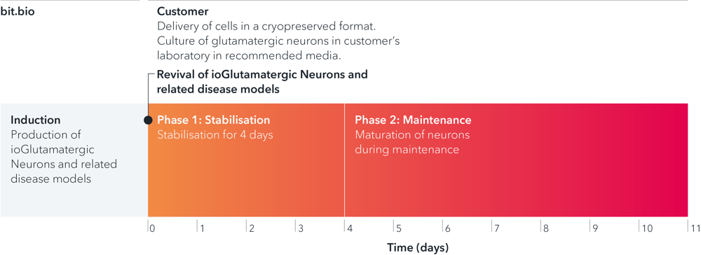
ioGlutamatergic Neurons MAPT [S305N;P301S]/[S305N;P301S] are delivered in a cryopreserved format and are programmed to mature rapidly upon revival in the recommended media. The protocol for the generation of these cells is a two-phase process: Phase 1, Stabilisation for 4 days; Phase 2, Maintenance, during which the neurons mature. Phases 1 and 2 after revival of cells are carried out by the customer.
Adult-like 4R:3R tau isoform expression
Cells carrying the S305N mutation express equimolar 4R:3R MAPT isoforms by day 11
MAPT isoform expression analysis by RT-qPCR, quantifying total, 3R and 4R MAPT expression in wild-type ioGlutamatergic Neurons (WT) and MAPT S305N disease model cells at days 11 and 32.
RNA (100 ng) was reverse-transcribed and amplified using TaqMan Fast Advanced Master Mix (Thermo Fisher Scientific) and isoform-specific probes (total MAPT: Hs00902194_m1; 3R MAPT: Hs00902192_m1, 4R MAPT: Hs00902312_m1). Expression levels were normalised to HMBS and the 4R:3R MAPT ratio was calculated.
View additional MAPT disease model product pages
ioGlutamatergic Neurons MAPT S305N/S305N (io1115)
ioGlutamatergic Neurons MAPT S305N/S305N+P301S/WT (io1116)
Western blot confirms the presence of 4R tau protein in MAPT S305N disease model cells
Total tau and 4R tau expression profiles in MAPT mutant ioGlutamatergic Neurons
Western blot analysis of total tau and 4R tau in wild-type (WT) vs. MAPT S305N/S305N (+P301S) ioGlutamatergic Neurons at days 11 and 32. The Western blot for total tau (A) shows the presence of the 4R and 3R isoforms in all MAPT mutant neurons whereas the WT control shows only 3R tau.
Cells were lysed in RIPA buffer with protease inhibitors; total protein was quantified via BCA Protein Assay Kit. Lysates were pre-treated with lambda phosphatase (3h at 30°C).
Cell lysates and a tau ladder positive control were resolved on a 4–20% Mini-PROTEAN® TGX Stain-Free™ gel (120V, 90 min) and transferred to a PVDF membrane; 5 μg protein was loaded.
Membranes were blocked (10 min) and incubated overnight at 4°C with primary antibodies: Anti-Tau antibody [TAU-5] (abcam #ab80579, 1:1000), Tau 4R (E7T4F) Rabbit Monoclonal Antibody (Cell Signaling Technology #79327, 1:1000), or for the loading control, Anti-GAPDH antibody [6C5] (abcam #ab8245, 1:5000). Signal was detected via ECL following secondary antibody incubation.
View additional MAPT disease model product pages
ioGlutamatergic Neurons MAPT S305N/S305N (io1115)
ioGlutamatergic Neurons MAPT S305N/S305N+P301S/WT (io1116)
MAPT S305N disease model cells show tau hyperphosphorylation
The S305N homozygous mutation drives tau hyperphosphorylation compared to the genetically matched wild-type control.
Western blot analysis of phospho-tau (p-tau) at days 11 and 32 in wild-type ioGlutamatergic Neurons (WT) and MAPT S305N disease model cells (A, B).
Cells were lysed in RIPA buffer with protease and phosphatase inhibitors; total protein was quantified via BCA Protein Assay Kit. Samples were resolved on a 4–20% Mini-PROTEAN® TGX Stain-Free™ gel (120V, 75 min) and transferred to a PVDF membrane; 10 μg protein was loaded.
Membranes were blocked (10 min) and incubated overnight at 4°C with primary antibodies: Phospho-Tau (Ser202, Thr205) Monoclonal Antibody (AT8) (Thermo Fisher #MN1020, 1:1000), Anti-Tau (phospho S396) antibody [E178] (abcam #ab32057, 1:1000), Anti-GAPDH antibody [6C5] (abcam #ab8245, 1:5000). Signal was detected via ECL following secondary antibody incubation.
Densitometry was performed relative to GAPDH signal using ImageJ and arbitrary units plotted (C, D).
View additional MAPT disease model product pages
ioGlutamatergic Neurons MAPT S305N/S305N (io1115)
ioGlutamatergic Neurons MAPT S305N/S305N+P301S/WT (io1116)
4R tau is absent in the wild-type and most abundant in the MAPT S305N/S305N disease model cells
Representative immunocytochemistry images of 4R tau expression in wild-type (WT) and MAPT S305N/S305N (+P301S) ioGlutamatergic Neurons at day 32.
Cells were fixed in 4% PFA and stained for the neuronal marker Beta-III tubulin (TUBB3) and 4R tau (Cell Signaling Technology #79327). Fluorescent signals were visualised using an ImageXpress microscope.
View additional MAPT disease model product pages
ioGlutamatergic Neurons MAPT S305N hom (io1115)
ioGlutamatergic Neurons MAPT S305N hom P301S het (io1116)
Highly characterised and defined
ioGlutamatergic Neurons MAPT [S305N;P301S]/[S305N;P301S] express neuron-specific markers comparably to the wild type control

Immunofluorescent staining on post-revival day 11 demonstrates similar homogenous expression of pan-neuronal proteins MAP2 and TUBB3 (upper panel) and glutamatergic neuron-specific transporter VGLUT2 (lower panel) in ioGlutamatergic Neurons MAPT [S305N;P301S]/[S305N;P301S] compared to the genetically matched control; (10x objective).
ioGlutamatergic Neurons MAPT [S305N;P301S]/[S305N;P301S] form structural neuronal networks by day 11

ioGlutamatergic Neurons MAPT [S305N;P301S]/[S305N;P301S] mature rapidly, show glutamatergic neuron morphology and form structural neuronal networks over 11 days, highly similar to the genetically matched control. Day 1 to 11 post thaw; (10x objective).
ioGlutamatergic Neurons MAPT [S305N;P301S]/[S305N;P301S] demonstrate gene expression of neuronal-specific and glutamatergic-specific markers following deterministic cell programming

Gene expression analysis demonstrates that ioGlutamatergic Neurons MAPT [S305N;P301S]/[S305N;P301S] and wild type ioGlutamatergic Neurons (WT Control) lack the expression of the pluripotency marker POU5F1 (OCT4) at day 11, while robustly expressing pan-neuronal (TUBB3 and SYP) and glutamatergic-specific (VGLUT1 and VGLUT2) markers. Gene expression levels were assessed by RT-qPCR (data normalised to HMBS; cDNA samples of the parental human iPSC line (hiPSC) were included as reference). Data represents day 11 post-revival samples, n=2 replicates.
View the step-by-step RNA extraction and RT-qPCR protocol used to generate this data
Industry leading seeding density

The recommended minimum seeding density is 30,000 cells/cm2, compared to up to 250,000 cells/cm2 for other similar products on the market. One small vial can plate a minimum of 0.7 x 24-well plate, 1 x 96-well plate, or 1.5 x 384-well plates. This means every vial goes further, enabling more experimental conditions and more repeats, resulting in more confidence in the data.
Adult-like 4R:3R tau isoform expression
Cells carrying the S305N mutation express equimolar 4R:3R MAPT isoforms by day 11
MAPT isoform expression analysis by RT-qPCR, quantifying total, 3R and 4R MAPT expression in wild-type ioGlutamatergic Neurons (WT) and MAPT S305N disease model cells at days 11 and 32.
RNA (100 ng) was reverse-transcribed and amplified using TaqMan Fast Advanced Master Mix (Thermo Fisher Scientific) and isoform-specific probes (total MAPT: Hs00902194_m1; 3R MAPT: Hs00902192_m1, 4R MAPT: Hs00902312_m1). Expression levels were normalised to HMBS and the 4R:3R MAPT ratio was calculated.
View additional MAPT disease model product pages
ioGlutamatergic Neurons MAPT S305N/S305N (io1115)
ioGlutamatergic Neurons MAPT S305N/S305N+P301S/WT (io1116)
Western blot confirms the presence of 4R tau protein in MAPT S305N disease model cells
Total tau and 4R tau expression profiles in MAPT mutant ioGlutamatergic Neurons
Western blot analysis of total tau and 4R tau in wild-type (WT) vs. MAPT S305N/S305N (+P301S) ioGlutamatergic Neurons at days 11 and 32. The Western blot for total tau (A) shows the presence of the 4R and 3R isoforms in all MAPT mutant neurons whereas the WT control shows only 3R tau.
Cells were lysed in RIPA buffer with protease inhibitors; total protein was quantified via BCA Protein Assay Kit. Lysates were pre-treated with lambda phosphatase (3h at 30°C).
Cell lysates and a tau ladder positive control were resolved on a 4–20% Mini-PROTEAN® TGX Stain-Free™ gel (120V, 90 min) and transferred to a PVDF membrane; 5 μg protein was loaded.
Membranes were blocked (10 min) and incubated overnight at 4°C with primary antibodies: Anti-Tau antibody [TAU-5] (abcam #ab80579, 1:1000), Tau 4R (E7T4F) Rabbit Monoclonal Antibody (Cell Signaling Technology #79327, 1:1000), or for the loading control, Anti-GAPDH antibody [6C5] (abcam #ab8245, 1:5000). Signal was detected via ECL following secondary antibody incubation.
View additional MAPT disease model product pages
ioGlutamatergic Neurons MAPT S305N/S305N (io1115)
ioGlutamatergic Neurons MAPT S305N/S305N+P301S/WT (io1116)
MAPT S305N disease model cells show tau hyperphosphorylation
The S305N homozygous mutation drives tau hyperphosphorylation compared to the genetically matched wild-type control.
Western blot analysis of phospho-tau (p-tau) at days 11 and 32 in wild-type ioGlutamatergic Neurons (WT) and MAPT S305N disease model cells (A, B).
Cells were lysed in RIPA buffer with protease and phosphatase inhibitors; total protein was quantified via BCA Protein Assay Kit. Samples were resolved on a 4–20% Mini-PROTEAN® TGX Stain-Free™ gel (120V, 75 min) and transferred to a PVDF membrane; 10 μg protein was loaded.
Membranes were blocked (10 min) and incubated overnight at 4°C with primary antibodies: Phospho-Tau (Ser202, Thr205) Monoclonal Antibody (AT8) (Thermo Fisher #MN1020, 1:1000), Anti-Tau (phospho S396) antibody [E178] (abcam #ab32057, 1:1000), Anti-GAPDH antibody [6C5] (abcam #ab8245, 1:5000). Signal was detected via ECL following secondary antibody incubation.
Densitometry was performed relative to GAPDH signal using ImageJ and arbitrary units plotted (C, D).
View additional MAPT disease model product pages
ioGlutamatergic Neurons MAPT S305N/S305N (io1115)
ioGlutamatergic Neurons MAPT S305N/S305N+P301S/WT (io1116)
4R tau is absent in the wild-type and most abundant in the MAPT S305N/S305N disease model cells
Representative immunocytochemistry images of 4R tau expression in wild-type (WT) and MAPT S305N/S305N (+P301S) ioGlutamatergic Neurons at day 32.
Cells were fixed in 4% PFA and stained for the neuronal marker Beta-III tubulin (TUBB3) and 4R tau (Cell Signaling Technology #79327). Fluorescent signals were visualised using an ImageXpress microscope.
View additional MAPT disease model product pages
ioGlutamatergic Neurons MAPT S305N hom (io1115)
ioGlutamatergic Neurons MAPT S305N hom P301S het (io1116)


Cell culture hacks | human iPSC-derived glutamatergic neurons
Read this blog on glutamatergic neuron cell culture for our top tips on careful handling, cell plating and media changes to achieve success from the outset.

Wild Type and Isogenic Disease Model cells: A true comparison
Further your disease research by pairing our wild type cells with isogenic disease models.


ioCells catalogue
Human iPSC-derived cells
powered by opti-ox
Consistent. Defined. Scalable.