Transfecting human iPSC-derived cells
Reliable transfection in human ioCells
Efficient transfection in human iPSC-derived cells depends on cell population, culture conditions and delivery methods suited to post-mitotic cells. Our ioCells are built to meet these needs, offering consistent, defined human cells alongside optimised transfection workflows. When gene delivery becomes predictable and reproducible, researchers can focus on generating biologically meaningful data rather than repeatedly iterating cell transfection protocols.
In addition, CRISPR-Ready ioCells and ioTracker Cells further streamline workflows by removing the need for transfection.

Consistent cells
Proven lot-to-lot consistency in ioCells supports reliable gene delivery and improved transfection efficiency.

Optimised protocols
Access transfection protocols specifically developed for hard-to-transfect, post-mitotic human cell types.

Defined cells
Every ioCells product is characterised by ICC, RT-qPCR and RNA-Seq, supporting predictable transfection outcomes.
ioCells in action
ioCells in action
High mRNA transfection efficiency
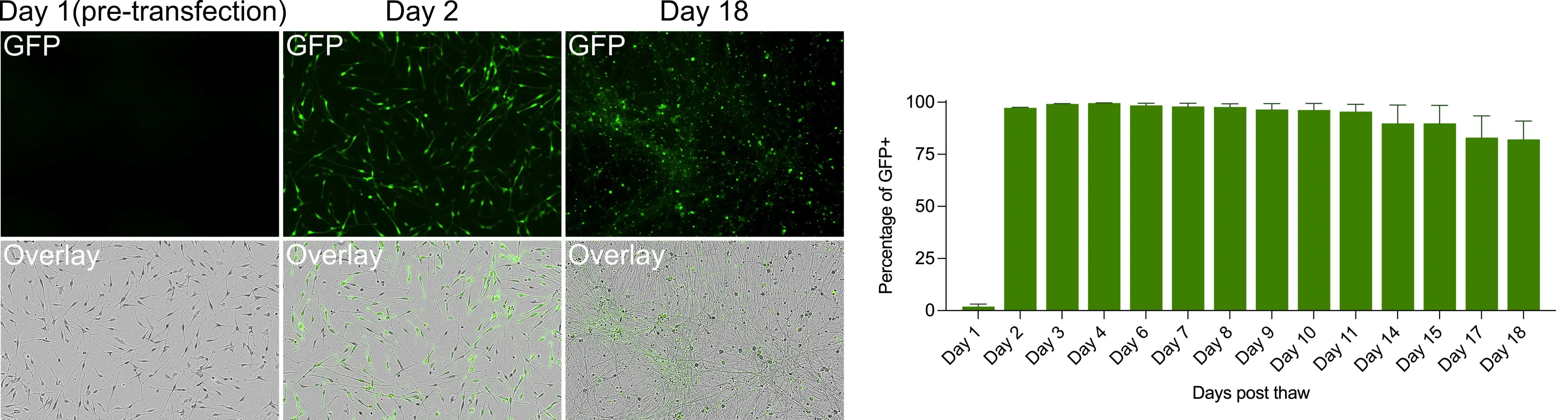
Fig 1. mRNA transfection in human glutamatergic neurons. ioGlutamatergic Neurons were transfected with EGFP mRNA and imaged over time to assess transfection efficiency. Cells seeded at 45,000 cells/cm² were transfected 24 h post-revival with 50 ng/well EGFP mRNA. Representative images show GFP expression on Days 1, 2, and 18, with quantification (right) confirming >95% GFP-positive cells and sustained expression up to 18 days.
ioGlutamatergic Neurons provide a consistent, reliable foundation that supports efficient mRNA delivery. Lipid-based transfection of EGFP mRNA in ioGlutamatergic Neurons yields high transfection efficiency, with stable expression maintained over time.
Post-transfection cell viability
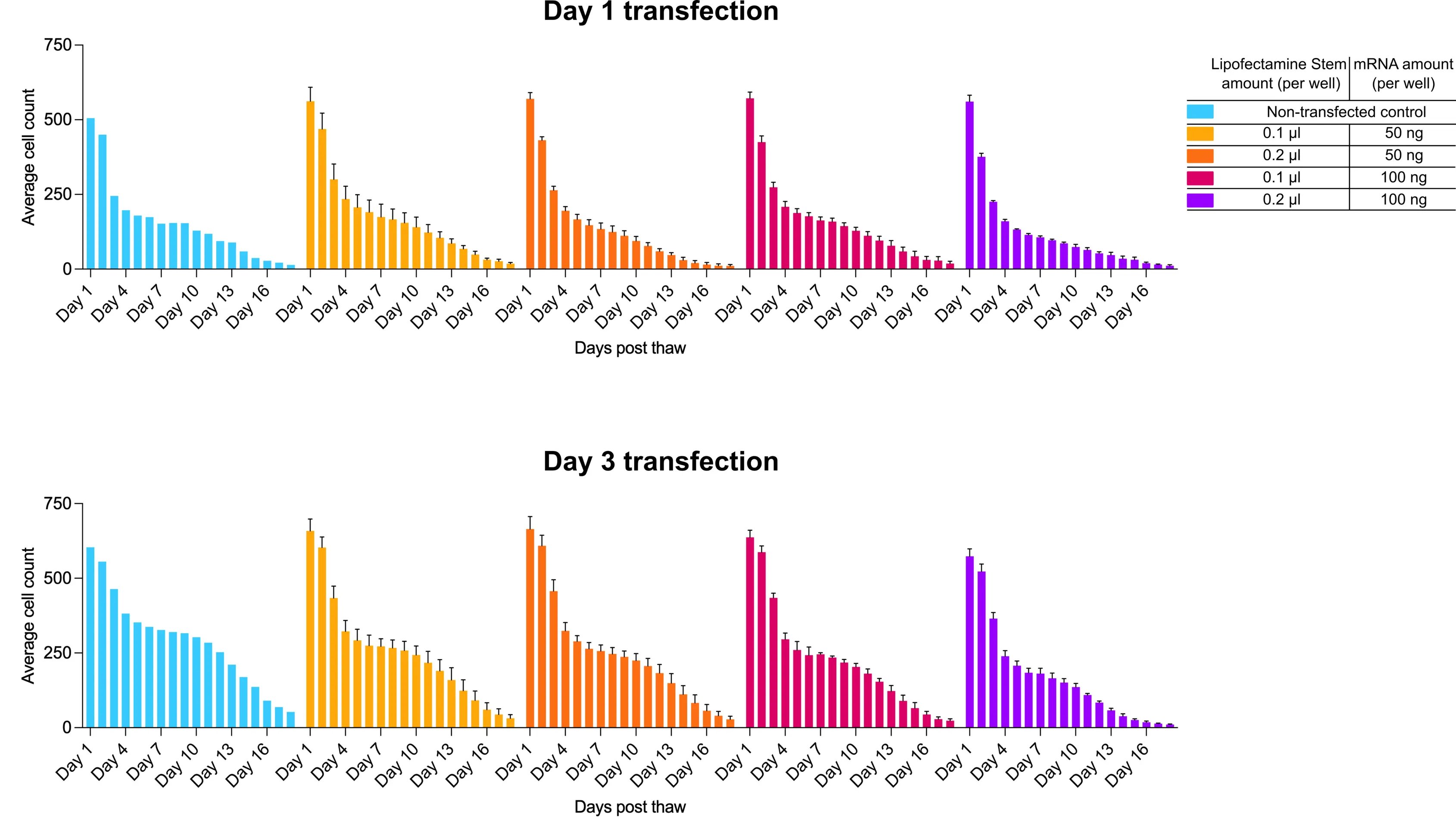
Fig 2. Cell viability following mRNA transfection in human motor neurons. ioMotor Neurons were transfected with EGFP mRNA using graded doses of Lipofectamine Stem and mRNA amount per well [control (blue); 0.1–0.2 µL reagent with 50–100 ng mRNA (others), respectively] at either Day 1 or Day 3 post-revival. Cell viability, measured by cell counts, remained comparable to non-transfected controls across all conditions, with marginally higher survival observed after Day 3 transfection.
Optimal transfection efficiency and viability depend on timing and mRNA/reagent ratios. In transfected ioMotor Neurons, cell viability data indicate negligible toxicity across different test conditions, with a slight viability benefit observed at day 3 post-neuron transfection compared to day 1.
Gene knockouts in human iPSC-derived microglia

Fig 3. Flow cytometry analysis of B2M protein expression in CRISPR-ready human microglia. At day 10 post-thaw, CRISPRko-Ready ioMicroglia were delivered a gRNA targeting B2M via lentiviral transduction or lipid-based transfection. After 5 days, B2M protein expression was assessed by flow cytometry. (A) Lentiviral delivery transduced ~20% of cells (GFP+), of which (B) a knockout of 86% was achieved, while (C) lipid-based transfection yielded 82% knockout.
CRISPR-Ready ioMicroglia facilitate rapid, robust gene editing in defined human cells with fewer transfection steps. After delivery of a gRNA targeting beta-2 microglobulin (B2M), flow cytometry data indicate high knockout efficiencies in both lentiviral delivery and lipid-based transfection.
Improving transduction efficiency
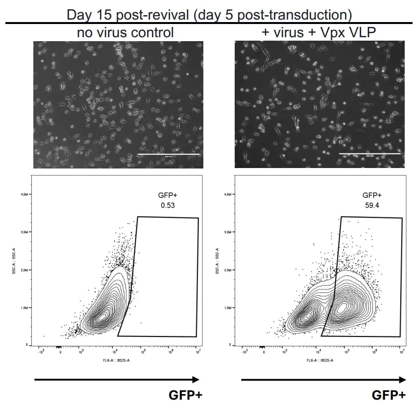
Fig 4. Lentiviral transduction of ioMicroglia. ioMicroglia cultured in a 6-well format at 80,000 cells/cm² were transduced at Day 10 post-thaw with GFP-containing lentivirus (1:100 dilution from a 20x concentrated prep) in the presence of Vpx VLPs. Five days post-transduction, no morphological differences were observed compared to control cells, and FACS analysis confirmed GFP+ cells in the majority of the population.
Lentiviral transduction of ioMicroglia, a cell type typically resistant to viral uptake, is enhanced by Vpx VLPs, which are virus-like particles engineered to package Vpx. This approach supports efficient genetic manipulation in human iPSC-derived microglia for functional and disease-focused studies.
Built-in fluorescence to track cells
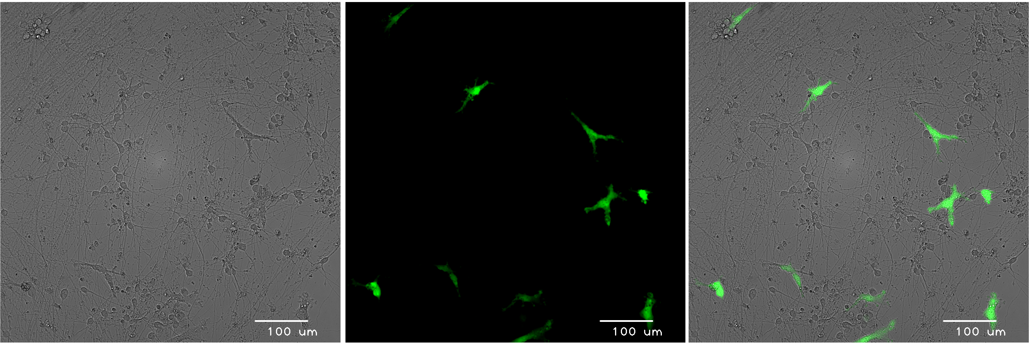
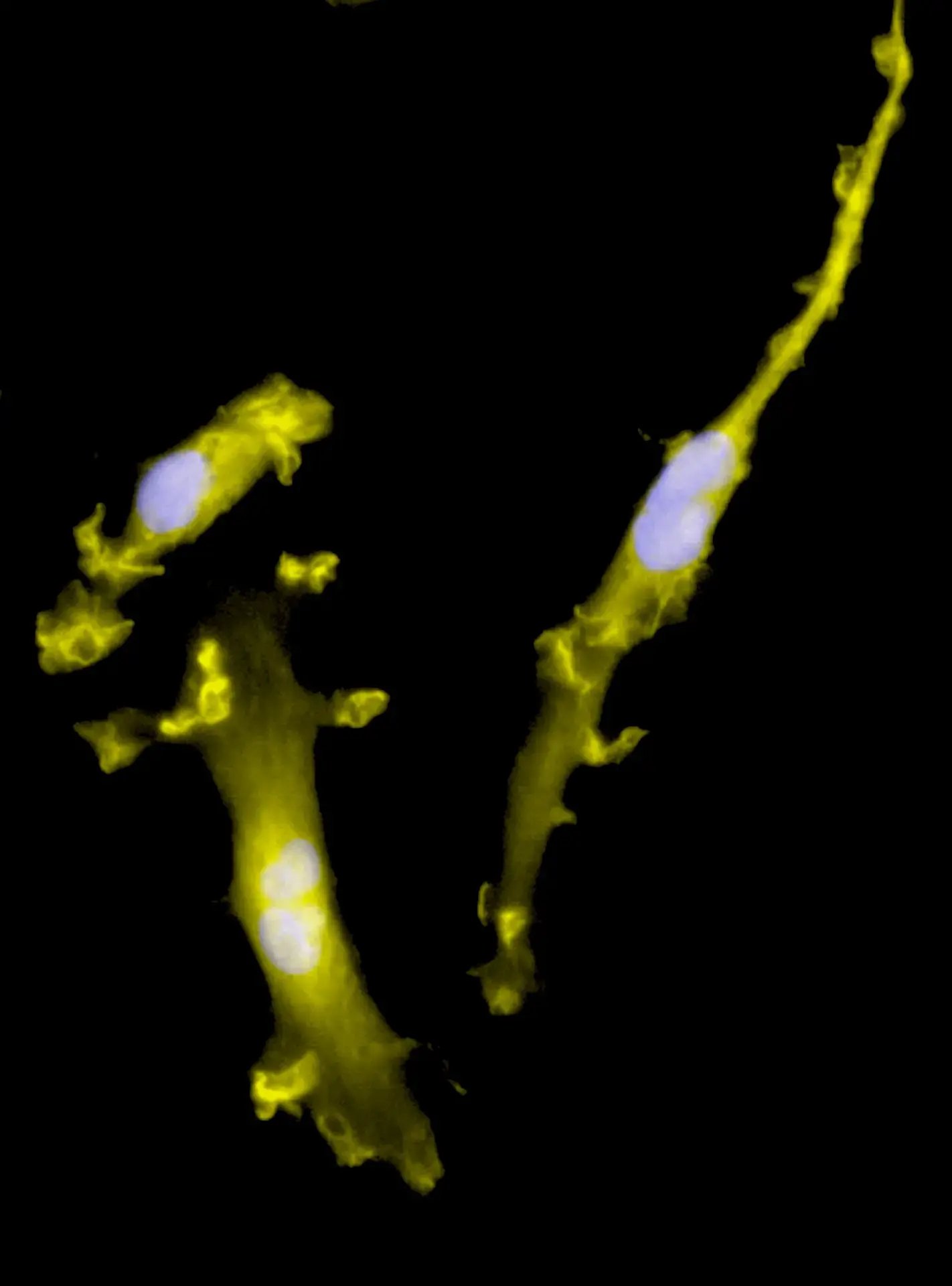
Fig 5. Live-cell imaging of human microglia in neuronal co-culture. ioGlutamatergic Neurons and ioTracker GFP ioMicroglia were cultured to day 10 post-thaw. On Day 11, the GFP ioMicroglia were directly added to the neurons, and the co-cultures were maintained for 3 additional days before live imaging. Brightfield and fluorescence images were acquired using a Leica DMi8 and merged, showing a distribution of GFP ioMicroglia within the co-culture.
ioTracker Cells – human iPSC-derived cells that constitutively express fluorescent proteins – enable live-cell imaging right from the outset. Visualise cell behaviour, track morphology, and examine cell-cell interactions in co-culture in real time, without additional labelling or reporter transfection.
Transfection tips from the bit.bio team

Tulin Tatar Ozkan, PhD
Senior Scientist II, bit.bio
“We have tested DNA transfection in ioCells using Lipofectamine Stem transfection reagent as well as several other commercially available transfection kits. However, achieving efficient DNA delivery into post-mitotic cells is very challenging; the transfection efficiency was typically below 1%. Therefore, we recommend using mRNA transfection, which consistently yields much higher transfection ef..."

Kaiser Karim, PhD
Scientist, bit.bio
“As neuronal cell bodies form connections across the culture plate via neurites, the disruption of one neuron can impact the entire culture. The key is to avoid causing a ‘tear’ in the culture when handling neurons. Tilt the cell culture plate during media changes to prevent cells from lifting off the surface.”
What scientists say about ioCells

Marijn Vlaming, PhD
Charles River Laboratories
“bit.bio cells do show very consistent phenotypes and there is not much batch to batch variation. They are delivered in high numbers of batches if we need them, and we can get them delivered very quickly.”
Recommended resources
Ready to browse?
Explore bit.bio’s portfolio of highly characterised, consistent, and physiologically relevant human iPSC-derived ioCells, designed to provide a strong foundation for transfection applications, and subsequently, reliable functional analyses.