cat no | io1096 Early Access
GFP ioMicroglia Male
Male human iPSC donor-derived microglia constitutively expressing GFP
-
Cryopreserved human iPSC-derived cells powered by opti-ox, that are ready for functional experiments in 4 days
-
Microglia constitutively expressing GFP, easy to track in multi-cellular cultures, ideal for live-cell imaging
-
Consistently perform key phagocytic and cytokine secretion functions, and are co-culture compatible
Male human iPSC donor-derived microglia constitutively expressing GFP
ioMicroglia Media Kit
Cell culture media kit for the culture of ioMicroglia up to 10 days post-thaw
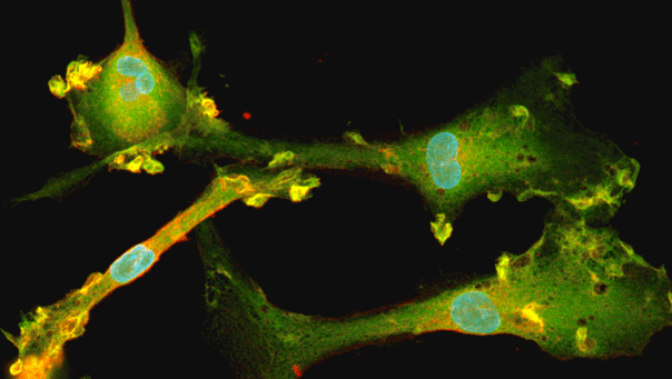
GFP ioMicroglia express key microglia markers
Immunofluorescent staining on day 10 post-revival demonstrates similar homogenous expression of microglia markers P2RY12 and IBA1 and ramified morphology in GFP ioMicroglia compared to ioMicroglia Male (io1021). GFP expression can be visualised throughout the cytosol in every cell for GFP ioMicroglia, but not in the WT control. 10X magnification.
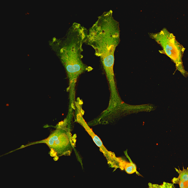
GFP ioMicroglia express key microglia markers
Immunofluorescent staining on day 10 post-revival demonstrates similar homogenous expression of microglia marker P2RY12 and ramified morphology in GFP ioMicroglia compared to ioMicroglia Male (io1021). GFP expression can be visualised throughout the cytosol in every cell for GFP ioMicroglia, but not in the WT control. 10X magnification.
Flow cytometry analysis of GFP expression at day 11 and day 21 for GFP ioMicroglia
Flow cytometry analysis demonstrating GFP expression in over 99.5% of cells for GFP ioMicroglia cultured until day 11, with no GFP expression seen in ioMicroglia Male (io1021). At day 21, the percentage of cells expressing GFP and the intensity does not decrease over time, indicating there is no silencing of the reporter gene.
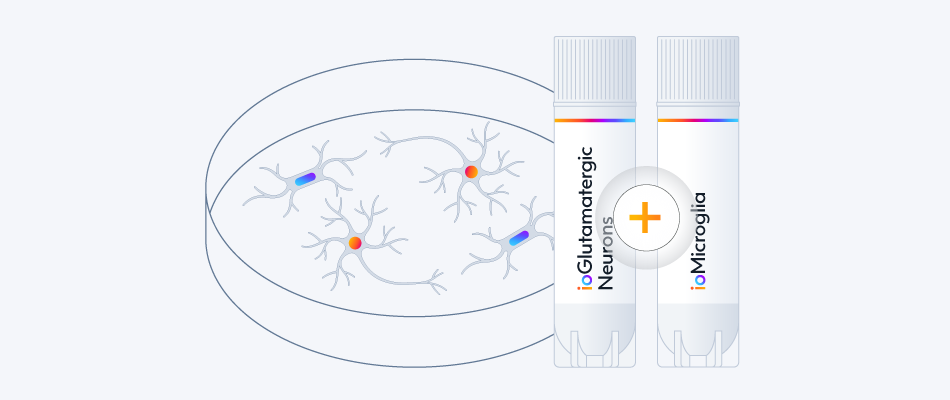
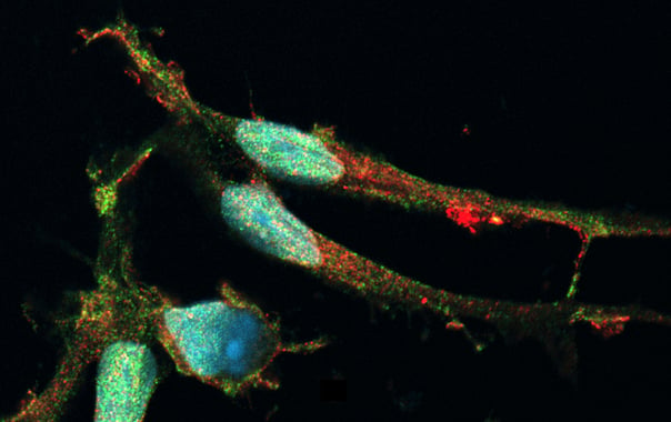
Live-cell imaging reveals clear visualisation of GFP ioMicroglia when co-cultured with ioGlutamatergic Neurons
ioGlutamatergic Neurons (io1001) were cultured to day 10 post-thaw. GFP ioMicroglia were cultured to day 10 post-thaw and were directly added to day 11 ioGlutamatergic Neurons. The co-cultures were maintained for a further 3 days before live-cell imaging with Leica DMi8. Brightfield and fluorescence images were taken and merged, easily demonstrating distribution of GFP ioMicroglia within the co-culture.
Easy-to-use co-culture protocol for GFP ioMicroglia with ioGlutamatergic Neurons
This co-culture protocol describes a method of co-culturing GFP ioMicroglia with ioGlutamatergic Neurons and associated disease models.
Phagocytosis of E. coli particles by GFP ioMicroglia
(A) Phagocytosis assay using pHrodo™ E. coli BioParticles™ at day 10 post-thaw demonstrates efficient uptake of bacteria particles by GFP ioMicroglia in comparison to ioMicroglia Male (io1021) +/- cytochalasin D control (an inhibitor of actin polymerisation). The graph displays that the proportion of cells phagocytosing E.coli particles over 24 hours for three technical replicates.
(B) The graph displays that the degree of cells phagocytosing E.coli particles over 24 hours. Images were acquired every 30 mins on the Incucyte® looking at red fluorescence and phase contrast. Three technical replicates were performed.
GFP ioMicroglia secrete pro-inflammatory cytokines upon activation
GFP ioMicroglia were stimulated at day 10 post-thaw with LPS 100 ng/ml and IFNɣ 20 ng/ml for 24 hours. Supernatants were harvested and analysed using MSD V-plex Proinflammatory Kit. GFP ioMicroglia secrete TNF⍺, IL-6, IL-8, IL-1b, IL-12p70 and IL-10 in response to the inflammatory stimuli. GFP ioMicroglia demonstrate a similar response to ioMicroglia Male (io1021), as expected. Three technical replicates were performed per lot.
View the stimulation for cytokine release protocol used to generate this data.
GFP ioMicroglia show ramified morphology by day 10
GFP ioMicroglia mature rapidly, and key ramified morphology can be identified by day 4 and continues through to day 10, similarly to ioMicroglia Male (io1021). Day 1 to 10 post-thawing; 100x magnification.
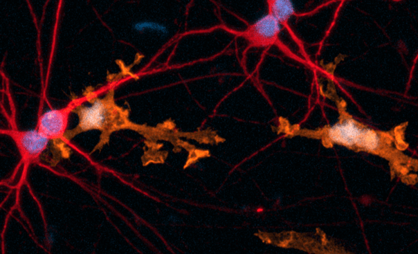
Phenotypic characterisation of a human iPSC-derived tri-culture using ioGlutamatergic Neurons, ioAstrocytes, and ioMicroglia
Using our fully optimised protocol, ioGlutamatergic Neurons (MAP2, red), ioMicroglia (IBA1, yellow) and ioAstrocytes (vimentin, cyan) were co-cultured to create a highly defined CNS model. High-resolution ICC analysis confirms the successful co-localisation and morphological health of three distinct cell types within a unified environment. By day 7, the protocol yields a highly consistent, integrated network suitable for complex cell modelling. DAPI (blue) highlights the total cell density and integrity of the culture. This protocol is compatible with derivative products of the three cell types, ensuring straightforward implementation across experimental workflows.
Vial limit exceeded
A maximum number of 20 vials applies. If you would like to order more than 20 vials, please contact us at orders@bit.bio.


Hoescht(blue)TUBB3(blue)_day4.png?width=604&name=bit.bio_ioGlutamatergic%20Neurons_60xMAP2(red)Hoescht(blue)TUBB3(blue)_day4.png)


_MAP2(R)_Tubb3(B)_Hoechst(B)_20x_merge-comp.jpg?width=604&name=Colour%20webinar%20with%20it-bio%20ioGlutamatergic%20Neurons_VGLUT2(G)_MAP2(R)_Tubb3(B)_Hoechst(B)_20x_merge-comp.jpg)