

cat no | io6015
ioMicroglia P2RY12 null/WT
Human iPSC-derived microglia model
-
Cryopreserved human iPSC-derived cells powered by opti-ox, that are ready for functional experiments in 4 days
-
Built to investigate the impact of P2RY12 knockout for neuroinflammation research
-
Consistently perform key phagocytic and cytokine secretion functions, and are co-culture compatible

Human iPSC-derived microglia for neuroinflammation research

Phagocytosis of ioMicroglia P2RY12null/wt is demonstrated comparably to the WT control and ioMicroglia P2RY12null/null
Phagocytosis was analysed at day 10 post-revival after incubation with 1 µg/0.33 cm2 pHrodo RED labelled E. coli particles for 24 hours +/- cytochalasin D control. The graph displays the proportion of cells phagocytosing E.coli over 24 hours. ioMicroglia P2RY12null/wt cells display a similar proportion of phagocytosis compared to the ioMicroglia P2RY12null/null and WT control. Images were acquired every 30 mins on the Incucyte® looking at red fluorescence and phase contrast. Three technical replicates were performed experiment.

Key cytokine secretion function displayed by ioMicroglia P2RY12 null/wt
Cytokine secretion was analysed at day 10 post-revival after stimulation with LPS 100 ng/ml and IFNɣ 20 ng/ml for 24 hours. This revealed that ioMicroglia P2RY12null/wt cells display a similar level of cytokine secretion compared to the ioMicroglia P2RY12null/null and WT control. Supernatants were harvested and analysed using MSD V-plex Proinflammatory Kit. Three technical replicates were performed per experiment.

ioMicroglia P2RY12 null/wt express IBA1 comparably to the genetically matched wild-type control
Immunofluorescent staining on day 10 post-revival demonstrates similar homogenous expression of the microglia marker IBA1 and ramified morphology in ioMicroglia P2RY12 null/wt cells compared to the genetically matched wild-type control, ioMicroglia Male. 100X magnification.

ioMicroglia P2RY12 null/wt show expected ramified morphology by day 10
ioMicroglia P2RY12null/wt cells mature rapidly and key ramified morphology can be identified by day 4 and continues through to day 10, similarly to the WT control. Day 1 to 10 post-thawing; 100x magnification.

P2RY12 null/wt heterozygous knockout confirmed by flow cytometry analysis
Flow cytometry analysis of ioMicroglia P2RY12 null/WT demonstrates heterozygous knockout of the P2RY12 gene translating to the protein level. Microglia purity demonstrated by >95% expression of CD45, CD11b and CD14 expression.
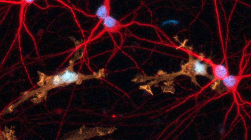
Female donor-derived ioMicroglia form co-cultures with ioGlutamatergic Neurons
ioGlutamatergic Neurons (io1001) were cultured to day 10 post-thaw. Female donor-derived ioMicroglia (io1029) cultured to either day 1 or day 10 post-thaw were added directly to day 10 ioGlutamatergic Neurons. The co-cultures were maintained for a further 6 days. Representative video showing that female donor-derived ioMicroglia form a stable co-culture with ioGlutamatergic Neurons. Live imaging was performed in 6.5-minute intervals over a time period of 3 hours and 31 minutes using the 3D Cell Explorer 96focus Nanolive Imaging system.

ioMicroglia are efficiently transfected with mRNA encoding GFP
ioMicroglia Male are efficiently transfected and show sustained long-term expression of mRNA encoding GFP. Cells were imaged throughout the experiment to assess transfection efficiency and evaluate potential cytotoxic effects of the transfection protocol. Day 4 images were captured prior to transfections on the same day.
Download the step-by-step protocol for lipid-based delivery of synthetic mRNA into ioMicroglia.
Vial limit exceeded
A maximum number of 20 vials applies. If you would like to order more than 20 vials, please contact us at orders@bit.bio.






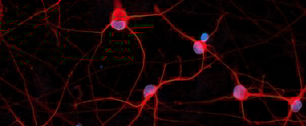
Hoescht(blue)TUBB3(blue)_day4.png?width=604&name=bit.bio_ioGlutamatergic%20Neurons_60xMAP2(red)Hoescht(blue)TUBB3(blue)_day4.png)






_MAP2(R)_Tubb3(B)_Hoechst(B)_20x_merge-comp.jpg?width=604&name=Colour%20webinar%20with%20it-bio%20ioGlutamatergic%20Neurons_VGLUT2(G)_MAP2(R)_Tubb3(B)_Hoechst(B)_20x_merge-comp.jpg)
Hoescht(blue)_day12v2.png?width=604&name=bit.bio_ioGlutamatergic%20Neurons_20xMAP2(red)Hoescht(blue)_day12v2.png)
