Co-culturing human iPSC-derived cells
Reproducible co-culture experiments rely on combining defined cell populations at optimised ratios. ioCells provide characterised, lot-consistent human cell inputs, supported by optimised protocols, helping researchers build controlled, robust co- and multi-culture models that deliver reliable experimental outcomes.

Defined cell inputs
Combine two or more of our ioCells with well-characterised, defined cell identities to build reliable, controllable co-culture or multi-culture systems.

Lot-to-lot consistency
Reduce inter-experimental variability and achieve reproducible co-culture outcomes using ioCells with proven lot-to-lot consistency.

Optimised protocols
Achieve scalable, defined co-cultures with optimised protocols offering guidance on seeding ratios, timing, media recipes, reagents, and more.
ioCells in action
ioCells in action
Enhanced network activity in co-cultures
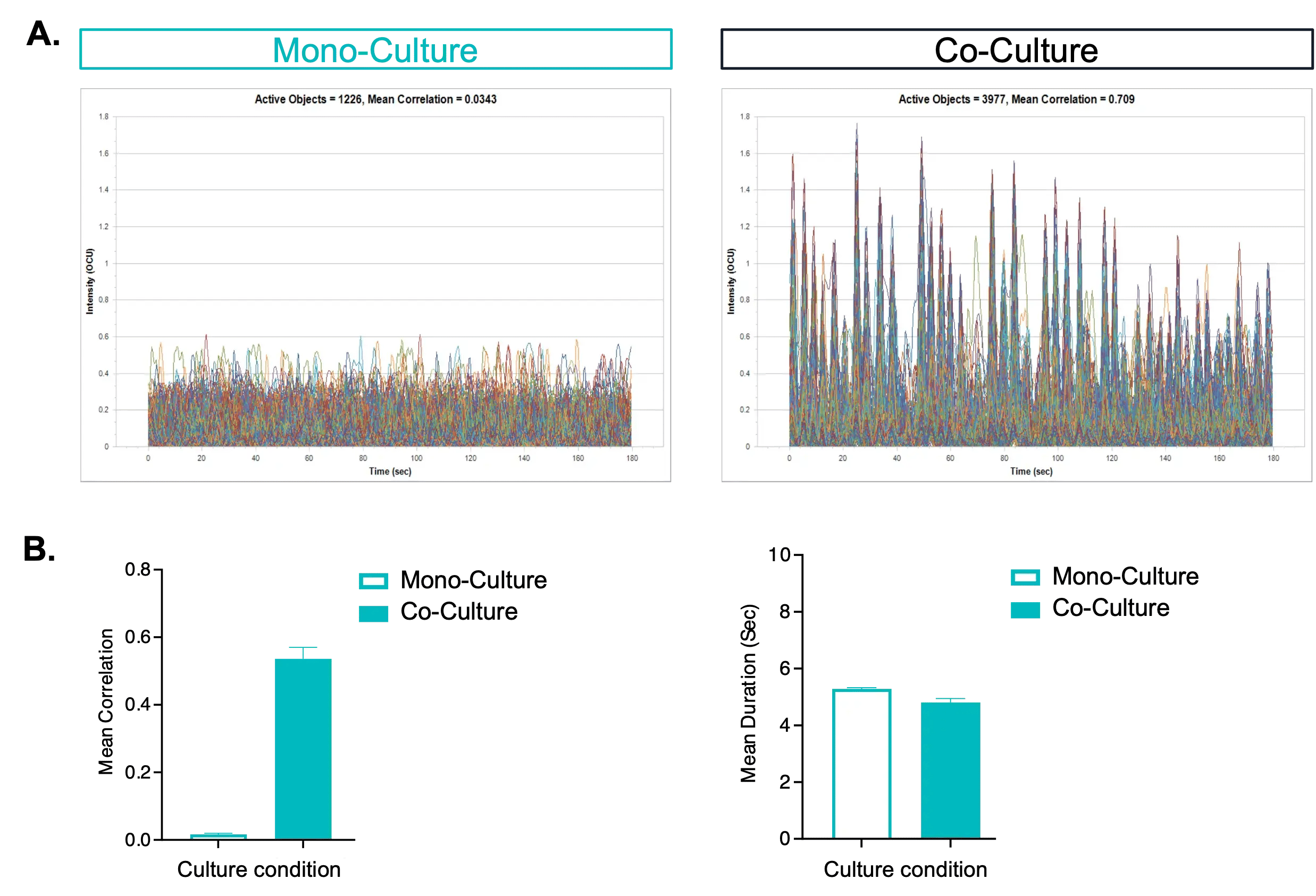
Fig 1. Measuring network activity in microglia-neuronal co-culture. (A) Representative calcium activity traces of ioGlutamatergic Neurons expressing Incucyte® Neuroburst Orange Lentivirus in monoculture or co-culture with ioMicroglia at day 15 post-addition. Monitored using the Incucyte® Neuronal Activity Analysis software. (B) Quantification shows increased mean network correlation in co-culture, with comparable mean burst duration (mean ± SEM, n = 3–12). Data courtesy of Sartorius.
Co-culturing ioMicroglia with ioGlutamatergic Neurons increases synchronised firing without altering burst duration. This demonstrates how defined cell co-culture systems can reveal microglial influence on neuronal activity, supporting studies of neuroinflammation and neuro-microglial network dynamics.
Tracking GFP ioMicroglia live
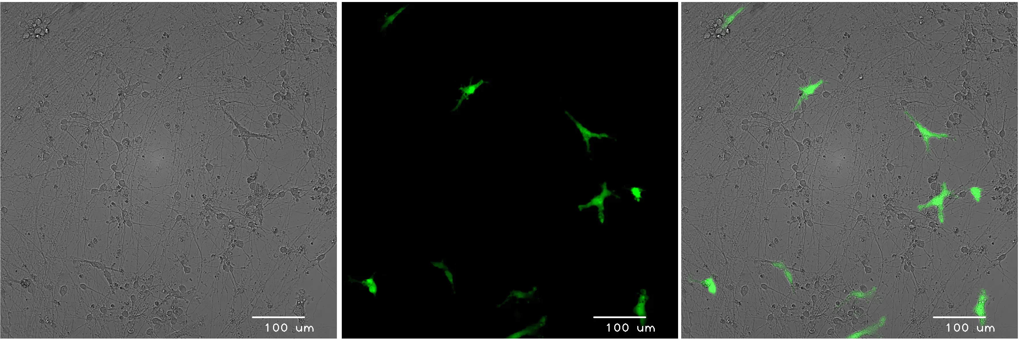
Fig 2. Live-cell imaging of GFP ioMicroglia in co-culture with ioGlutamatergic Neurons. ioGlutamatergic Neurons were cultured to day 10 post-thaw. GFP ioMicroglia, cultured to day 10 post-thaw, were added directly to day 11 ioGlutamatergic Neurons. Co-cultures were maintained for a further 3 days before live-cell imaging on a Leica DMi8.
Constitutive GFP expression allows for the tracking of ioMicroglia within a neuronal network without antibody staining. This supports live-cell imaging of microglial morphology, motility and activation state, and simplifies cell sorting in multi-cell cultures.
Co-culture with ioSkeletal Myocytes
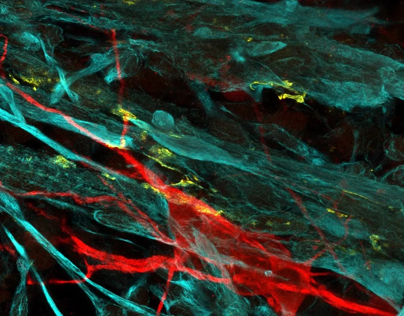
Fig 3. Confocal imaging of motor neuron–skeletal muscle co-culture. ioMotor Neurons were co-cultured with ioSkeletal Myocytes and analysed by confocal microscopy at day 30. Alpha-bungarotoxin (yellow) staining highlights acetylcholine receptor expression on co-cultured ioSkeletal Myocytes. Meanwhile, desmin (cyan) and microtubule-associated protein 2 (red) identify ioSkeletal Myocytes and ioMotor Neurons, respectively. (40X magnification.)
In co-cultures of ioMotor Neurons and ioSkeletal Myocytes, acetylcholine receptor expression indicates early neuromuscular junction-like organisation. This co-culture method provides a foundation for studying neuromuscular junction formation and related cellular processes.
GABAergic modulation of network activity

Fig 4. MEA analysis of glutamatergic, GABAergic, and astrocyte tri-cultures. ioGlutamatergic Neurons and human iPSC-derived astrocytes were cultured alone or with increasing numbers of ioGABAergic Neurons and analysed on a 48-well Axion Maestro Pro MEA platform. (A) Network burst frequency increases in excitatory cultures and decreases with the addition of inhibitory neurons. (B) Active electrode counts show sustained spontaneous activity across conditions. Data courtesy of Charles River Laboratories.
ioGABAergic Neurons integrate into excitatory co-cultures to form functional networks and modulate overall network activity. This tri-culture system facilitates the modelling of excitatory–inhibitory interplay in vitro, enabling further study of circuit dynamics relevant to epilepsy and neurodegeneration.
Neuro-glial quad-culture systems
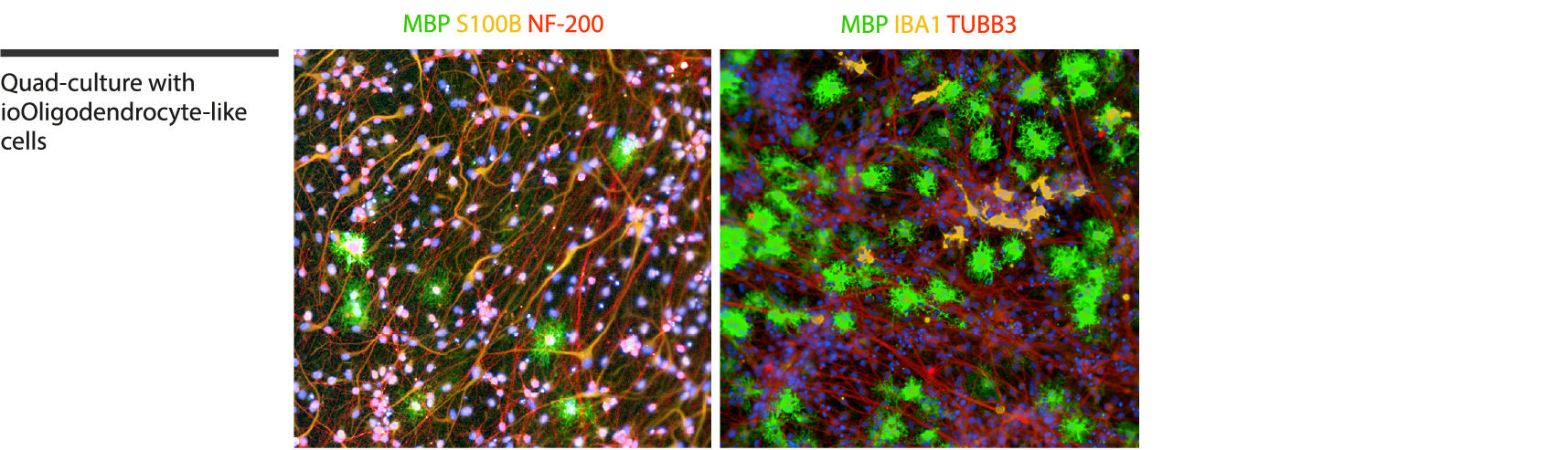
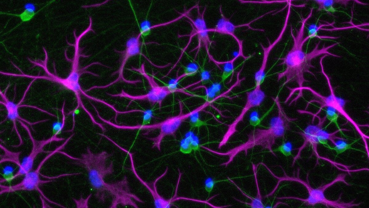
Fig 5. Immunofluorescent characterisation of a quad-culture model. Representative immunostaining shows ioOligodendrocyte-like cells expressing MBP (green), ioGlutamatergic Neurons expressing NF-200 (red) and TUBB3 (red), ioMicroglia expressing IBA1 (yellow), human iPSC-derived astrocytes expressing S100B (yellow), and DAPI counterstain (blue). Cultures were analysed on day 14.
By combining neurons, oligodendrocyte-like cells, microglia, and astrocytes, this quad-culture demonstrates successful integration of multiple human CNS cell types into a single in vitro system, offering a platform to study neurodegenerative processes relevant to Alzheimer’s disease and multiple sclerosis.
Taking multi-cell cultures into 3D
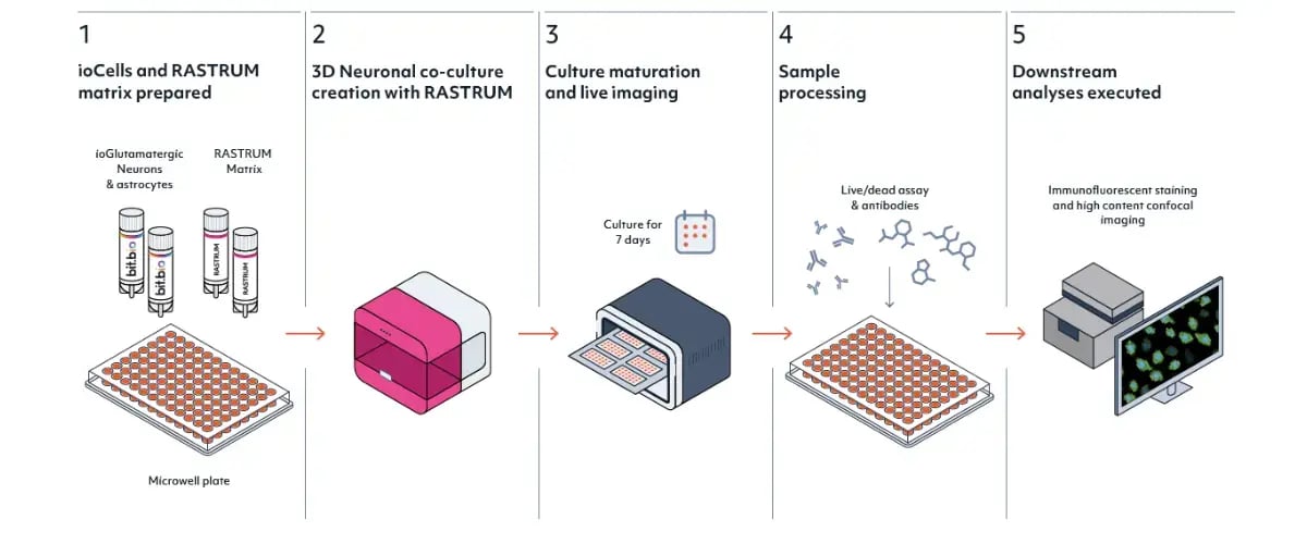
Fig 6. Automated generation of human neuronal microtissues. ioGlutamatergic Neurons and iPSC-derived astrocytes were bioprinted on the Inventia RASTRUM™ platform to form 3D co-cultures in a CNS-mimetic hydrogel, supporting high-throughput screening in 96- and 384-well formats. Data courtesy of Inventia Life Science.
Consistent inputs matter even more as model architecture grows. ioCells have been bioprinted into reproducible 3D neuronal microtissues and integrated into BrainZell brain organoids, where ioMicroglia add measurable inflammatory signalling, cytokine release and pharmacological modulation.
Optimised ioCells combinations
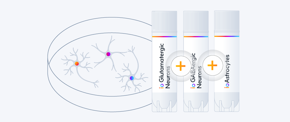
Start from one of our verified ioCells combinations, from neuron–glia co-cultures to neuromuscular and tri-cultures, then extend it with our optimisation guide. Each protocol gives step-by-step instructions, media recipes and recommended readouts.
Working with a combination we have not verified yet? The multi-cell culture protocols and optimisation guide covers coating, plating strategy, seeding densities, media transitions and validation of functional crosstalk.
Recommended resources
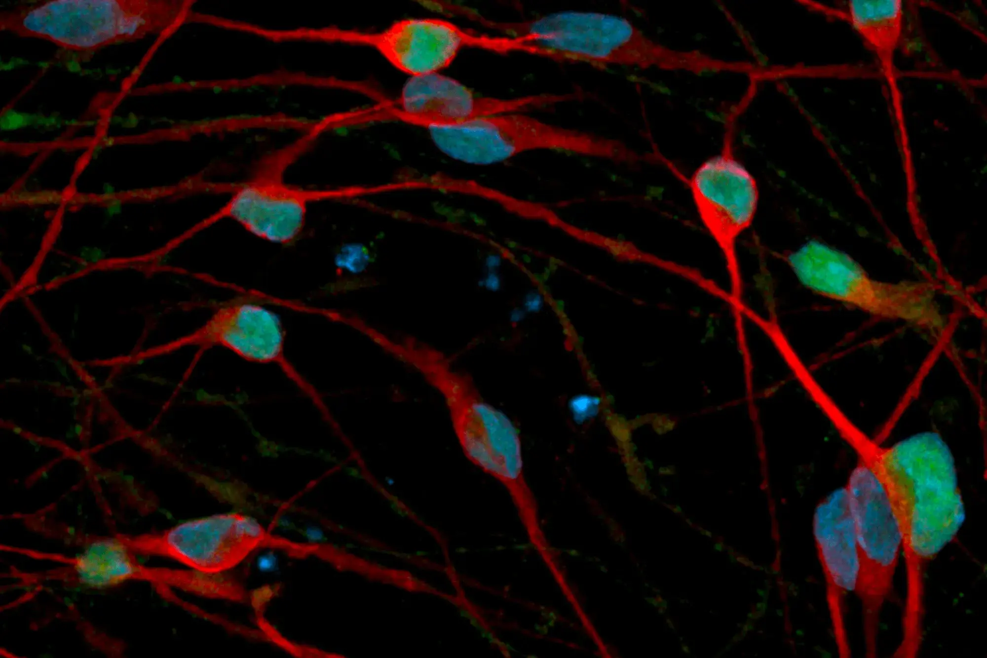
Ready to browse?
Explore our full portfolio of human iPSC-derived cells. From neurons and glia to muscle and immune cell types, our products are consistent and defined, supporting reliable data generation for applications in disease research and drug discovery.