cat no | io1022
ioGlutamatergic Neurons GBA null/N409S
Human iPSC-derived Gaucher and Parkinson’s disease model
-
Cryopreserved human iPSC-derived cells powered by opti-ox that are ready for experiments in days
-
Engineered to carry a GBA mutation relevant for modelling Gaucher and Parkinson's diseases
-
Consistent, functional excitatory neurons that form neuronal networks within days
Human iPSC-derived Gaucher and Parkinson’s disease model
Representative ICC staining of ioGlutamatergic Neurons GBA null/N409S; MAP2 (red), VGLUT2 (green), DAPI (blue).
ioGlutamatergic Neurons Media Kit
Cell culture media kit for the culture of ioGlutamatergic Neurons up to 14 days post-thaw
Reduced glucocerebrosidase activity demonstrated in disease model cells carrying GBA mutations
Glucocerebrosidase (GCase) activity was measured in ioGlutamatergic Neurons carrying mutations in the GBA gene (GBA null/R159W, GBA null/null and GBA null/N409S) or carrying SNCA A53T mutations (SNCA A53T/A53T and SNCA A53T/WT), and the wild-type genetically matched control.
Cells were seeded in 96-well plates (25,000 cells per well) and a fluorometric assay using 4-MUG as substrate was carried out on DIV9 to measure the enzymatic activity of GCase.
The assay confirmed that iPSC-derived glutamatergic neurons with GBA mutations showed significantly reduced GCase activity compared to the wild-type control and SNCA A53T disease models.
Statistical analysis: One-way ANOVA followed by Dunnett’s multiple comparison test versus isogenic control ***p<0.001.
Data courtesy of T. Loeffler et al., Scantox
ioGlutamatergic Neurons GBA null/N409S express neuron-specific markers comparably to the wild-type control
Immunofluorescent staining on day 11 post-thaw demonstrates similar homogenous expression of pan-neuronal proteins TUBB3 and MAP2 (upper panel) and glutamatergic neuron-specific transporter VGLUT2 (lower panel) in ioGlutamatergic Neurons GBA null/N409S compared to the genetically matched control (10X objective).
ioGlutamatergic Neurons GBA null/N409S form structural neuronal networks by day 11
ioGlutamatergic Neurons GBA null/N409S mature rapidly and form structural neuronal networks over 11 days, when compared to the genetically matched control. Day 1 to 11 post thaw; 10X objective.
Advanced tri-culture of ioGlutamatergic Neurons, ioAstrocytes 2.0, and ioMicroglia
While neuronal-astrocyte co-cultures capture essential structural and metabolic synergy, the addition of microglia, the resident myeloid cells of the CNS, is critical for accurately modelling neuroinflammatory pathways, synaptic pruning, and homeostatic maintenance.
This representative immunocytochemistry image demonstrates a successful tri-culture established at a 2:1:2 ratio (neurons:astrocytes:microglia). Using an optimised bit.bio protocol for 24-well plates, the tri-culture can be robustly maintained for up to 7 days. Staining with cell type-specific markers confirms the presence and integration of all three populations, shown alongside single-channel panels: MAP2-positive ioGlutamatergic Neurons (red), S100B-positive ioAstrocytes 2.0 (cyan), and IBA1-positive ioMicroglia (yellow); with DAPI as a nuclear counterstain (blue). Scale bars: 200 µm.
Efficient mRNA transfection into ioGlutamatergic Neurons
ioGlutamatergic Neurons are efficiently transfected and show sustained long-term expression of mRNA encoding GFP. ioGlutamatergic Neurons were imaged from day 1 post-thaw and throughout the experiment to assess transfection efficiency and evaluate potential cytotoxic effects of the transfection protocol. Day 1 images were captured prior to transfection on the same day.
Download the step-by-step protocol for lipid-based delivery of synthetic mRNA into ioGlutamatergic Neurons.
Lipid-based delivery of synthetic mRNA into ioGlutamatergic Neurons
ioGlutamatergic Neurons were transfected 24 hours post-thaw using Lipofectamine™ Stem Transfection Reagent. The transfection efficiency was evaluated by fluorescence imaging over 18 days after mRNA delivery, resulting in high transfection efficiency (close to 100%) and long-term sustained GFP expression.
Quantification of the GFP signal shows a decrease in GFP intensity over time, while the percentage of GFP-positive cells remains largely unchanged over time.
(A) The percentage of GFP-positive cells from two independent experiments.
(B) GFP intensity, quantified in successfully transfected cells from two independent experiments is quantified and normalised to day 2 (24 hours post-transfection).
Vial limit exceeded
A maximum number of 20 vials applies. If you would like to order more than 20 vials, please contact us at orders@bit.bio.
Benchtop benefits

Make true comparisons
Pair the ioDisease Model Cells with the genetically matched wild-type control to investigate the impact of the GBA mutation on molecular mechanisms and cell function.

Scalable
With opti-ox technology, we can make billions of consistently programmed cells, surpassing the demands of industrial workflows.

Quick
The disease model cells and wil-type control are experiment ready as early as 2 days post revival, and form structural neuronal networks at 11 days.
Cells arrive ready to plate
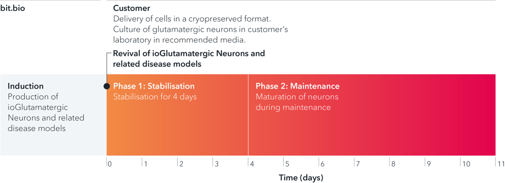
ioGlutamatergic Neurons GBA null/N409S are delivered in a cryopreserved format and are programmed to mature rapidly upon revival in the recommended media. The protocol for the generation of these cells is a two-phase process: Phase 1, Stabilisation for 4 days; Phase 2, Maintenance, during which the neurons mature. Phases 1 and 2 after revival of cells are carried out by the customer.
Disease-related phenotype
Reduced glucocerebrosidase activity demonstrated in disease model cells carrying GBA mutations
Glucocerebrosidase (GCase) activity was measured in ioGlutamatergic Neurons carrying mutations in the GBA gene (GBA null/R159W, GBA null/null and GBA null/N409S) or carrying SNCA A53T mutations (SNCA A53T/A53T and SNCA A53T/WT), and the wild-type genetically matched control.
Cells were seeded in 96-well plates (25,000 cells per well) and a fluorimetric assay using 4-MUG as substrate was carried out on DIV9 to measure the enzymatic activity of GCase.
The assay confirmed that iPSC-derived glutamatergic neurons with GBA mutations showed significantly reduced GCase activity compared to the wild-type control and SNCA A53T disease models.
Statistical analysis: One-way ANOVA followed by Dunnett’s multiple comparison test versus isogenic control ***p<0.001.
Data courtesy of T. Loeffler et al., Scantox
Highly characterised and defined
ioGlutamatergic Neurons GBA null/N409S express neuron-specific markers comparably to the wild-type control
Immunofluorescent staining on day 11 post-thaw demonstrates similar homogenous expression of pan-neuronal proteins TUBB3 and MAP2 (upper panel) and glutamatergic neuron-specific transporter VGLUT2 (lower panel) in ioGlutamatergic Neurons GBA null/N409S compared to the genetically matched control (10X objective).
ioGlutamatergic Neurons GBA null/N409S form structural neuronal networks by day 11
ioGlutamatergic Neurons GBA null/N409S mature rapidly and form structural neuronal networks over 11 days, when compared to the genetically matched control. Day 1 to 11 post thaw; 10X objective.
Industry leading seeding density

The recommended minimum seeding density is 30,000 cells/cm2, compared to up to 250,000 cells/cm2 for other similar commercially available products. One small vial can plate a minimum of 0.7 x 24-well plate, 1 x 96-well plate, or 1.5 x 384-well plates. This means every vial goes further, enabling more experimental conditions and more repeats, resulting in more confidence in the data.


Cell culture hacks | human iPSC-derived glutamatergic neurons
Read this blog on glutamatergic neuron cell culture for our top tips on careful handling, cell plating and media changes to achieve success from the outset.

Wild Type and Isogenic Disease Model cells: A true comparison.
Further your disease research by pairing our wild type cells with isogenic disease models.


ioCells catalogue
Human iPSC-derived cells
powered by opti-ox
Consistent. Defined. Scalable.