cat no | io1100 Early Access
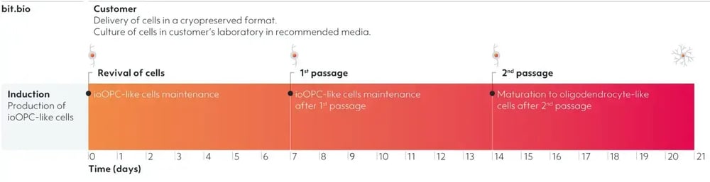
ioOPC-like cells
Human iPSC-derived OPC-like cells
- Cryopreserved human iPSC-derived cells powered by opti-ox, that are ready for experiments in days
- Ideal for proliferation assays for demyelinating disease
- Resemble human OPC phenotype and mature to oligodendrocyte-like cells
Human iPSC-derived OPC-like cells
ioOPC-like cells have limited proliferative ability and can be passaged
Assessment of cell fold increase in ioOPC-like cells after treatment with Click-iT™ EdU cell proliferation assay from ThermoFisher.
(A) Cell expansion during the OPC-maintenance phases, over 7-day periods, shows the cell number increasing by approximately 4- to 5-fold. n=3 biological replicates.
(B) Quantification of EdU incorporation at day 9 (2 days post-1st passage) shows approximately 80% positive cells. n=3 biological replicates.
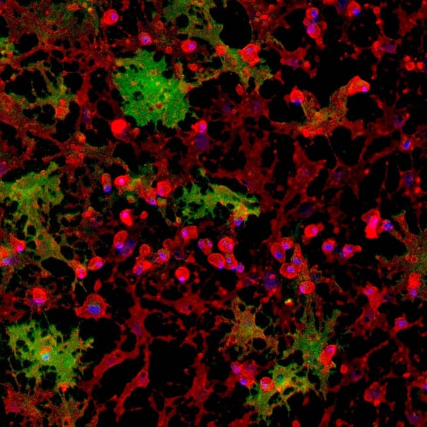
(C) Immunostaining imaging of the cells after treatment of Click-iT EdU
at day 9 (2 days post-1st passage). Cells are positive for O4 (red), the EdU marker (green), and the DAPI counterstain (blue).
View the step-by-step EdU proliferation assay protocol used to generate this data
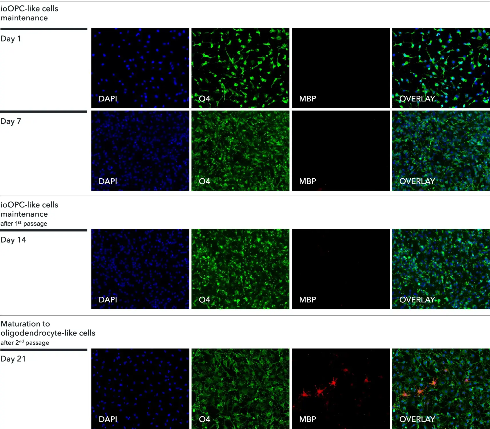
ioOPC-like cells express oligodendroglial-specific markers
Immunocytochemistry staining of cells shows expression of key markers. At day 1, 7 and 14, the cells are positive for the oligodendrocyte-specific marker O4 (green), and the DAPI counterstain (blue).
By day 21, after the 2nd passage, and upon switching to maturation media, cells show an increased complexity and are positive for O4 (green), the myelin basic protein (MBP) (red), and the DAPI counterstain (blue). Images acquired on the Echo Revolve microscope (10X objective).
View the step-by-step immunofluorescent staining protocol used to generate this data
Cells display an OPC-like morphology during maintenance phases, whilst keeping the ability to mature towards mature oligodendrocyte-like cells
Upon deterministic programming, cells show rapid morphological changes, during the different phases of culture, from maintenance to maturation. From day 1 to day 7, the cells display an OPC-like morphology. After the 1st passage, from day 8 to day 14, the cells continue to display an OPC-like morphology. After the 2nd passage, the cells mature rapidly and by day 21 display an oligodendrocyte-like morphology. Images acquired on the Incucyte® (10X objective).
Key oligodendroglial genes are expressed by ioOPC-like cells
Following deterministic programming, the cells downregulate expression of the pluripotency gene OCT4. The cells show robust expression of relevant oligodendroglial markers, including PDGFRA, GSPG4, CNP, PLP1, and MBP. Gene expression levels assessed by RT-qPCR, data expressed relative to the reference (housekeeping) gene, HMBS. Data represents day 1, 7, 14, and 21 post-revival samples; n=3 technical replicates.
View the step-by-step RNA extraction and RT-qPCR protocol used to generate this data
Bulk RNA-sequencing demonstrates expression of key oligodendroglial genes
ioOPC-like cells express key oligodendroglial genes as demonstrated on this heatmap generated from bulk RNA-sequencing analysis of the cells at multiple timepoints (day 0, day 7, day 14 and day 21).
During OPC maintenance phases, on day 7 and day 14, the cell population express oligodendrocyte progenitor genes, such as PDGFRA. Whilst on day 21, during the maturation phase, there is an increase in the expression of genes associated with mature oligodendrocytes, such as MBP, indicating that the cell population has matured towards an oligodendrocyte-like identity. Day 21 data was generated with 1 μM db-cAMP rather than the recently optimised medium formulation containing 100 μM db-cAMP (refer to this technical note to learn more).
A maximum number of 20 vials applies. If you would like to order more than 20 vials, please contact us at orders@bit.bio.


Hoescht(blue)TUBB3(blue)_day4.png?width=604&name=bit.bio_ioGlutamatergic%20Neurons_60xMAP2(red)Hoescht(blue)TUBB3(blue)_day4.png)