cat no | io1028
ioOligodendrocyte-like cells
Human iPSC-derived oligodendrocyte-like cells
- Cryopreserved human iPSC-derived cells powered by opti-ox, that are ready for experiments in days
- Ideal for compound screening and phenotypic assays for neurodegenerative and demyelinating disease
- Resemble a pre-myelinating oligodendrocyte and show increased MBP expression in co-culture with neurons
Human iPSC-derived oligodendrocyte-like cells
Generation of experimental readouts within 8 days post-revival
These cells are programmed to rapidly mature when in a monolayer culture. By following a simple protocol, these cells allow scientists to generate experimental readouts within 8 to 10 days post-thaw.
Enhancement of MBP expression by optimising db-cAMP concentration in media
Immunofluorescent staining of the cells at day 10 demonstrated a dose-dependent effect of db-cAMP in MBP expression and culture stability.
At 1 µM db-cAMP, only a subset of cells exhibit complex branching. In contrast, in cultures with 100 µM db-cAMP, a higher number of cells had a more mature phenotype, displaying complex and highly branched processes, and expressing MBP. On day 10, the proportion of MBP-expressing cells in the 100 µM db-cAMP condition increased approximately 4 to 5-fold compared to the 1 µM condition, reaching approximately 25% of the total number of cells in culture. All data was generated with a seeding density of 27,000 cells/cm2.
Dive into the complete dataset in the technical note.
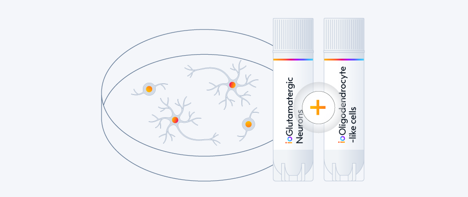
Long-term maintenance and myelination marker expression in ioGlutamatergic Neurons and ioOligodendrocyte-like cells co-culture
ioGlutamatergic Neurons and ioOligodendrocyte-like cells were co-cultured in media containing 1 µM or 100 µM db-cAMP and fixed at timepoints day 14 and 21. Immunocytochemistry was performed for MBP (green) and TUBB3 (red). Nuclei were counterstained with DAPI (blue). Images were acquired using a Zeiss LSM 900 confocal microscope (scale bar: 50 µm).
Representative confocal images illustrate potential neuronal–oligodendrocyte interactions and sustained myelination marker expression.
View the complete dataset and the step-by-step co-culture protocol
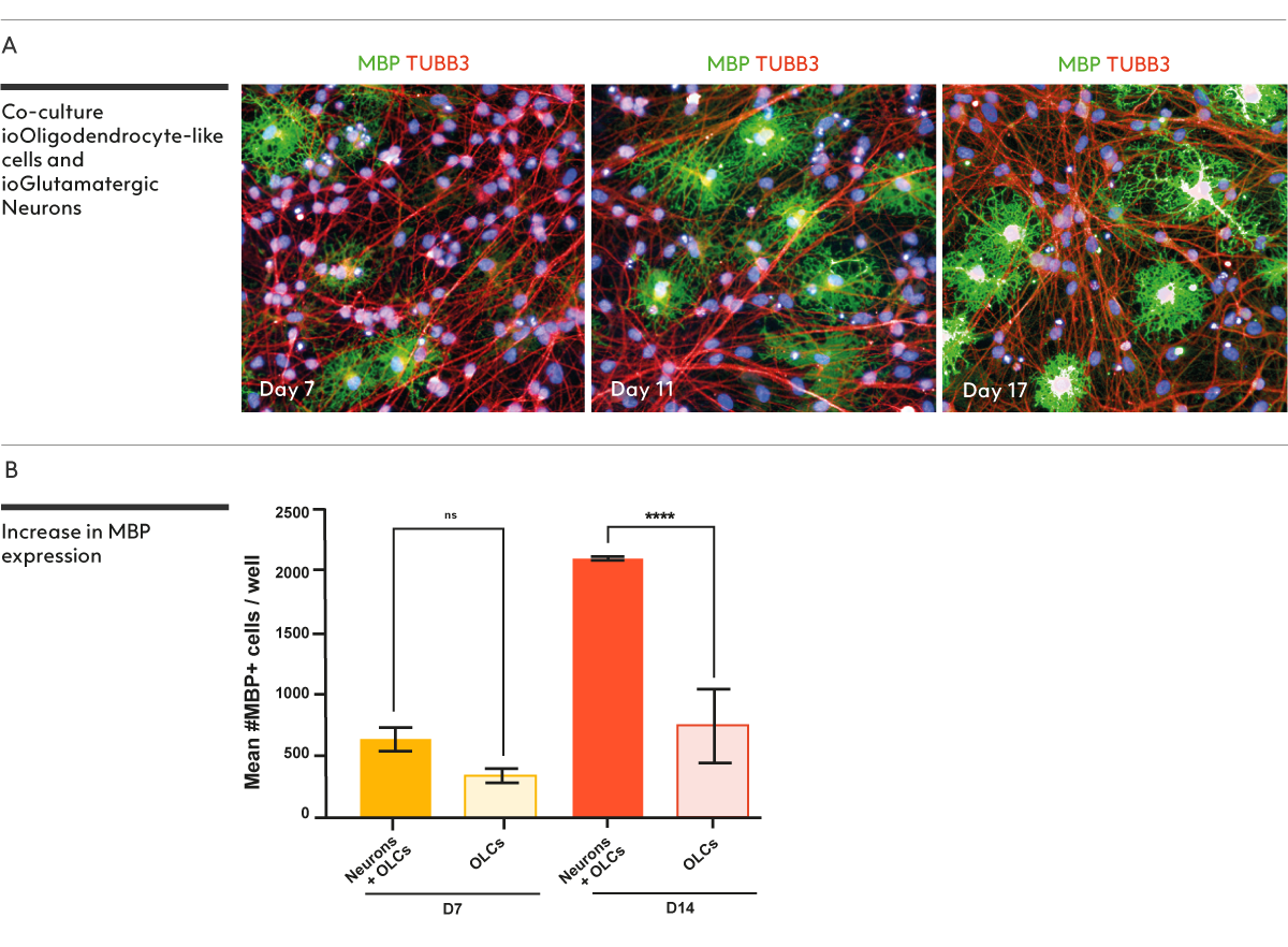
ioOligodendrocyte-like cells show increased MBP expression when in co-culture with ioGlutamatergic Neurons
The enhancement of MBP expression in a co-culture model provides a valuable in vitro cellular model for scientists studying neurons and oligodendrocytes interactions
(A) Immunofluorescent staining of a co-culture of ioOligodendrocyte-like cells expressing the oligodendrocyte marker MBP (green) and ioGlutamatergic Neurons pan-neuronal marker TUBB3 (red), and DAPI counterstain (blue). Co-cultures were analysed on day 7, day 11 and day 17.
(B) Graph shows that the co-culture of ioOligodendrocyte-like cells (represented as OLCs) with ioGlutamatergic Neurons (represented as Neurons) increases the number of MBP-positive cells over time (day 14 vs day 7) relative to ioOligodendrocyte-like cells mono-culture. High-content imaging and a custom algorithm were used to quantify the number of MBP-positive cells. n=2 technical replicates; One-way ANOVA with Tukey’s multiple comparison or unpaired T-test; *p<0.05; **p<0.005; ****p<0.0001; not-significant not indicated.
Data courtesy of Bsibsi, M. et al., 2024, Charles River Laboratories.
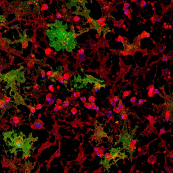
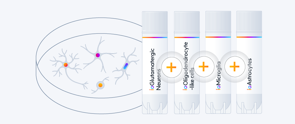
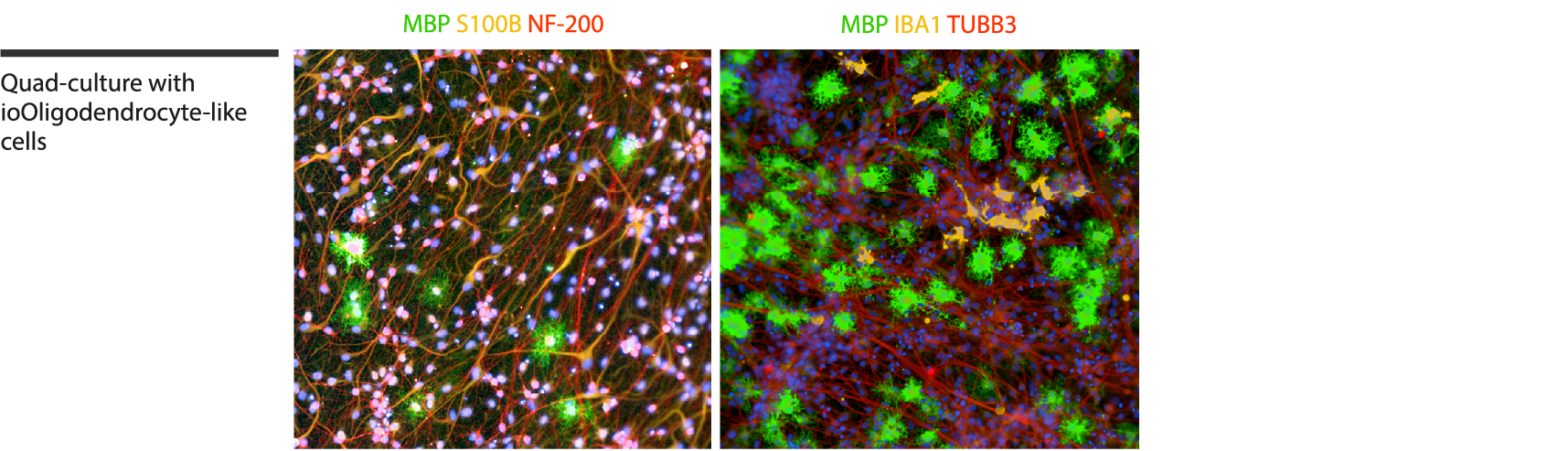
Quad-culture model with ioOligodendrocyte-like cells, ioGlutamatergic Neurons, ioMicroglia and human iPSC-derived astrocytes
Complex multi-cellular models have the potential to provide insights into the role of glial cells in disease mechanisms of neurodegenerative diseases, such as Alzheimer’s disease and multiple sclerosis.
Immunofluorescent staining of a multi-cellular culture including ioOligodendrocyte-like cells expressing MBP (green), ioGlutamatergic Neurons expressing NF-200 (red) and TUBB3 (red), ioMicroglia expressing IBA1 (yellow), human iPSC-derived astrocytes expressing S100B (yellow), and DAPI counterstain (blue). Cultures were analysed on day 14.
Data courtesy of Bsibsi, M. et al., 2024, Charles River Laboratories, presented in a poster at the Society of Neuroscience 2024 meeting.
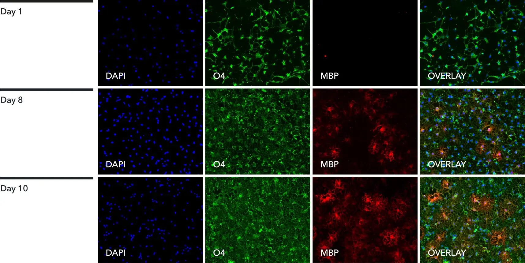
ioOligodendrocyte-like cells express oligodendroglial-specific markers
Immunofluorescent staining of the cells at day 1 (upper panel), day 8 (middle panel) and day 10 (lower panel) post-revival. At day 1, the cells are positive for the oligodendrocyte-specific marker O4 (green), and the DAPI counterstain (blue). At day 8 and 10, ioOligodendrocyte-like cells show an increased complexity and are positive for O4 (green), the myelin basic protein (MBP) (red), and the DAPI counterstain (blue). 100X magnification.
View the step-by-step immunofluorescent staining protocol used to generate this data
ioOligodendrocyte-like cells constitute a highly pure population of oligodendroglial cells
Single cell RNA-sequencing analysis was performed on ioOligodendrocyte-like cells at two timepoints: day 1 and day 8; iPSCs represent the parental non-induced human iPSC line. The cell population does not express astrocyte marker SOX9, subtype-specific astrocyte marker GFAP, nor microglia markers AIF (gene encoding for IBA1 protein) and TREM2. There is limited expression of neuronal marker RBFOX3. These data suggest that ioOligodendrocyte-like cells constitute a highly pure population of oligodendroglial cells. Gene expression was assessed by Parse Biosciences single cell RNA-sequencing. Data generated with 1 µM db-cAMP and a seeding density of 27,000 cells/cm2.
Cells show an oligodendrocyte-like morphology by day 8
Upon deterministic programming, cells show rapid morphological changes, acquiring an OPC-like morphology by day 1 post-revival. By day 8, cells have matured and display an oligodendrocyte-like morphology. Brightfield images show day 1, 3, 8, and 10 post-thawing; scale bar: 400 μm.
Key oligodendroglial genes are expressed by ioOligodendrocyte-like cells
Following deterministic programming, the cells downregulate expression of the pluripotency gene OCT4, whilst demonstrating robust expression of relevant oligodendroglial markers, including PDGFRA, MBP, MAG, and PLP1. Gene expression levels assessed by RT-qPCR, data expressed relative to the reference (housekeeping) gene, HMBS. Data represents day 1, 8 and 10 post-revival samples; n=3 technical replicates.
View the step-by-step RNA extraction and RT-qPCR protocol used to generate this data
Single cell RNA-sequencing shows ioOligodendrocyte-like cells express typical oligodendroglial markers and display increased maturity by day 8
Single cell RNA-sequencing analysis was performed on ioOligodendrocyte-like cells at two timepoints: day 1 and day 8; iPSCs represent the parental non-induced human iPSC line. By day 1, the cell population exhibits a distinct expression profile characteristic of oligodendroglial cells, demonstrated by the expression of oligodendrocyte progenitor genes such as PDGFRA and PTPRZ1. By day 8, there is an increase in the expression of genes associated with mature oligodendrocytes, such as MBP, PLP1, CNP, MYRF and MAL, indicating that the cell population has matured towards an oligodendrocyte-like identity. Gene expression was assessed by Parse Biosciences single cell RNA-sequencing. Data generated with 1 µM db-cAMP and a seeding density of 27,000 cells/cm2.
Whole transcriptome analysis demonstrates high lot-to-lot consistency of ioOligodendrocyte-like cells
Bulk RNA-sequencing analysis was performed on three different lots of manufactured product at day 1 and day 8 post revival. Principal component analysis (PCA) represents the variance in gene expression between the three different lots of ioOligodendrocyte-like cells. This analysis shows lots clustering very closely which demonstrates high consistency at each given timepoint. This lot-to-lot consistency in every vial gives scientists confidence in their experimental reproducibility. Colours represent the parental non-induced hiPSC cell line and the three lots of ioOligodendrocyte-like cells; shapes represent different timepoints. Data generated with 1 µM db-cAMP and a seeding density of 27,000 cells/cm2.
Expression levels for specific genes of interest can be requested by contacting our team at technical@bit.bio.
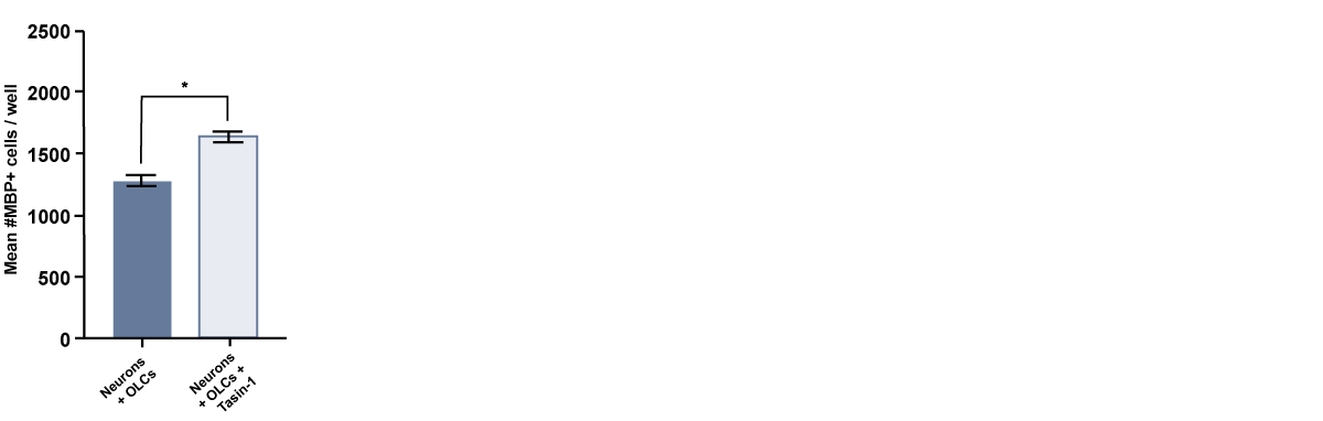
Treatment with Tasin-1 leads to increase in MBP expression
The enhancement in MBP expression in response to compound treatment indicates that the cells can be used for compound screening in early stage drug discovery workflows.
Treatment of ioOligodendrocyte-like cells with Tasin-1, a small molecule inhibitor of cholesterol biosynthesis enzymes and described as a pro-myelinating compound by Hubler et al, 2018, resulted in a significant increase in the number of MBP-positive cells. MBP quantification was performed using high-content imaging and a custom-developed algorithm. Timepoint day 14; n=2 technical replicates; One-way ANOVA with Tukey’s multiple comparison or unpaired T-test; *p<0.05; **p<0.005; ****p<0.0001; not-significant not indicated.
Data courtesy of Bsibsi, M. et al., 2024, Charles River Laboratories.
A maximum number of 20 vials applies. If you would like to order more than 20 vials, please contact us at orders@bit.bio.


.png?width=1715&height=396&name=ioOligodendrocyte-PCA_AllSamples2%20(1).png)


Hoescht(blue)TUBB3(blue)_day4.png?width=604&name=bit.bio_ioGlutamatergic%20Neurons_60xMAP2(red)Hoescht(blue)TUBB3(blue)_day4.png)