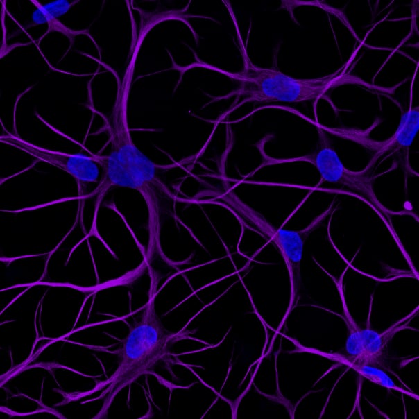
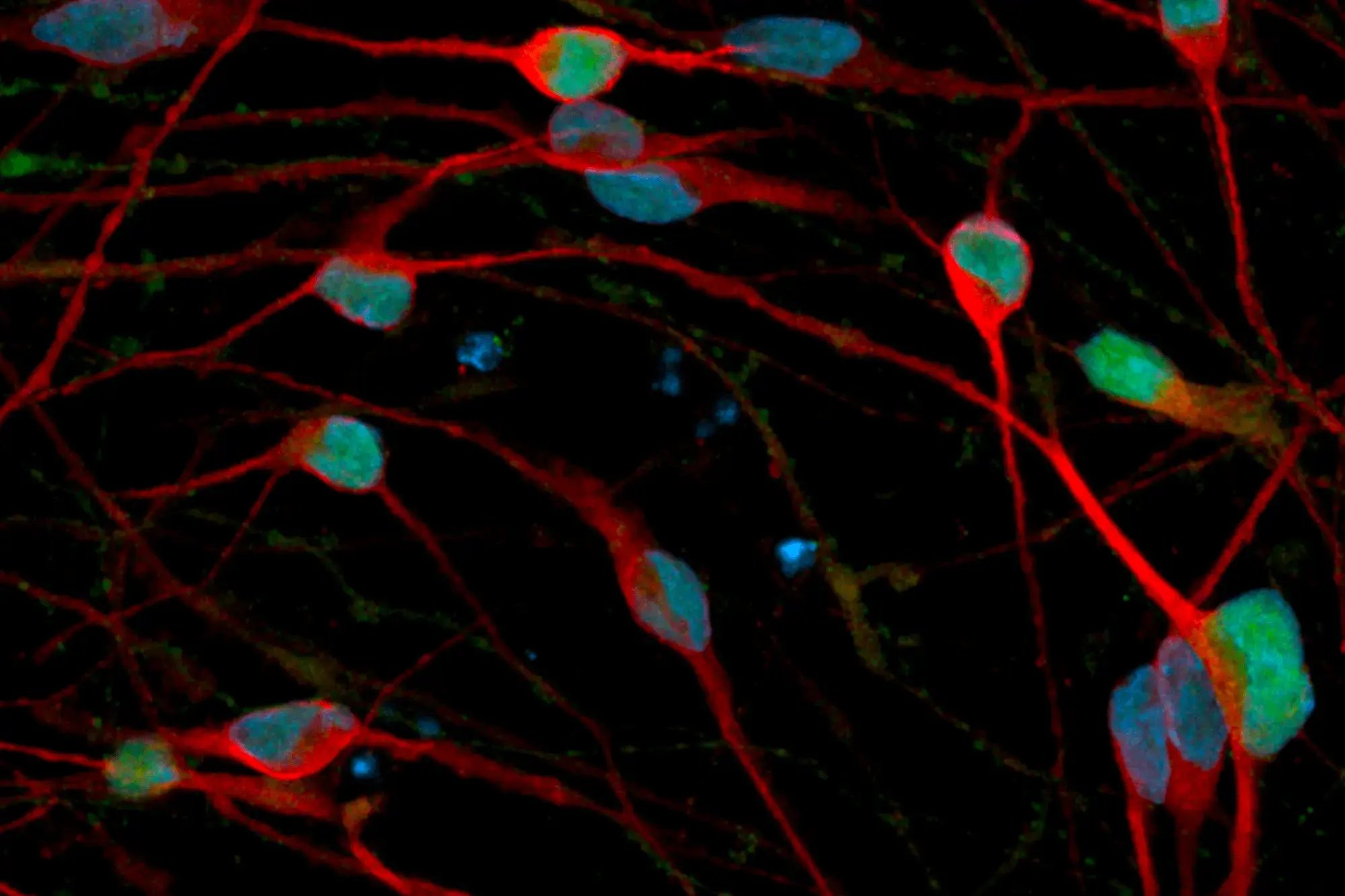
(UPDATED)ioAstrocytes_2.0_Hero_image_VIM_DAPI
(UPDATED)ioAstrocytes_2.0_ioGlutamatergic-Neurons_co-culture_MEA-DIV50
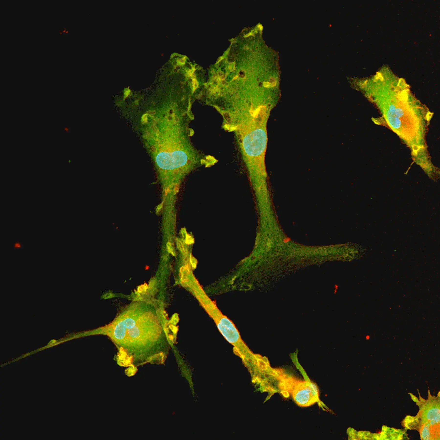
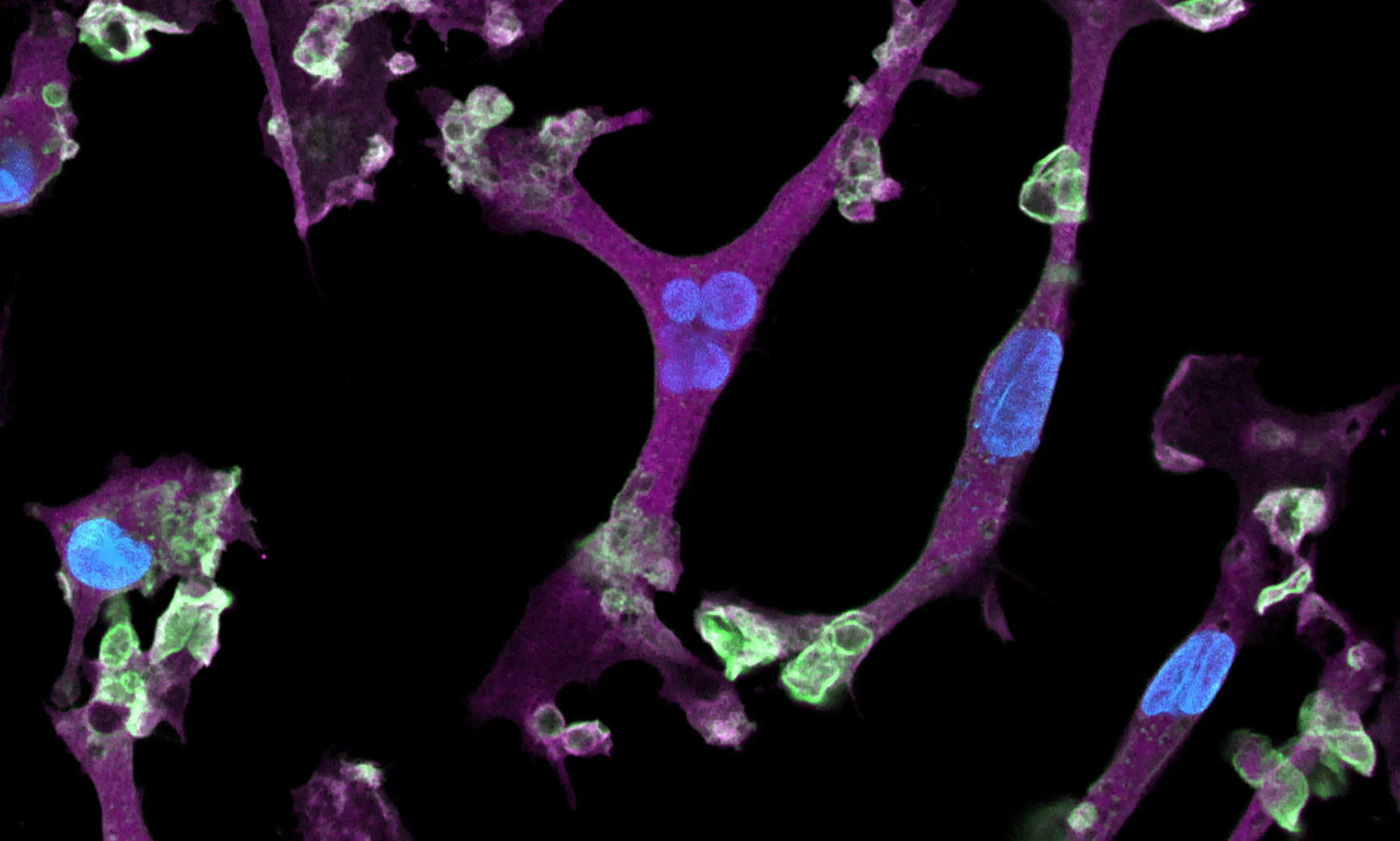
(UPDATED)ioAstrocytes_2.0_Phagocytosis_s.aureus_D7_thumbnail
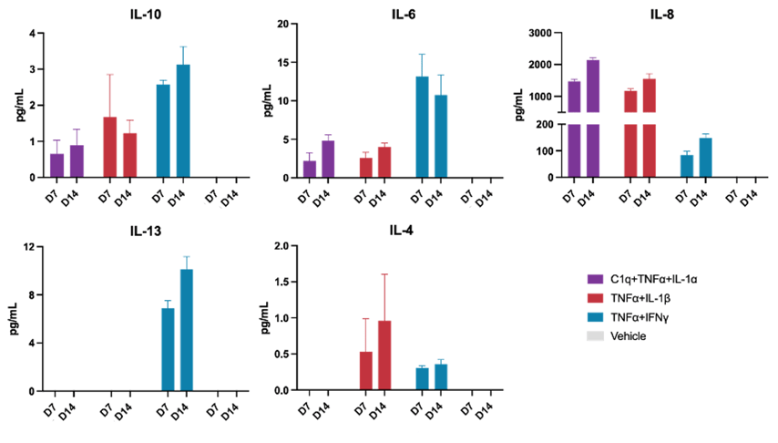
ioAstrocytes_2.0_cytokine-secretion_MSD_panel1
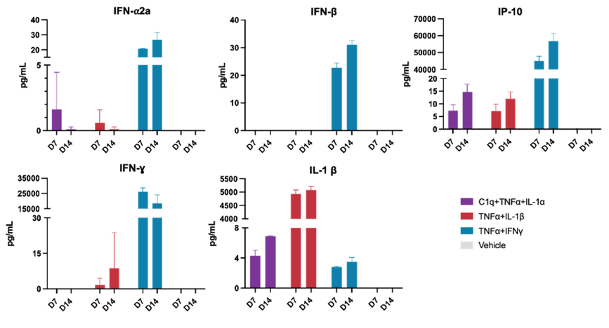
ioAstrocytes_2.0_cytokine-secretion_MSD_panel2
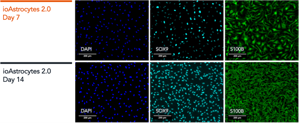
ioAstrocytes_2.0_ICC_Panel-1_DAPI_SOX9_S100B_D7_D14-1
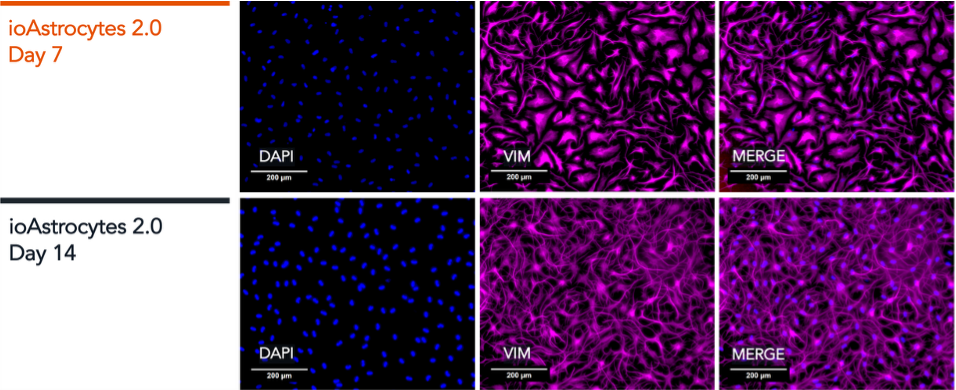
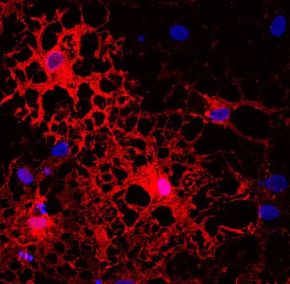
ioAstrocytes_2.0_ICC_Panel-2_DAPI_VIM_MERGE_D7_D14-1
ioAstrocytes_2.0_RT-qPCR_NANOG_OCT4_EAAT1_VIM_SOX9_S100B_iPSC_D1_D7_D14
ioAstrocytes_ioGlutamatergic-Neurons_co-culture_DIV13
Phenotypic analysis of a tri-culture for CNS modelling using ioGlutamatergic Neurons, ioAstrocytes, and ioMicroglia
(UPDATED)ioAstrocytes_2.0_Hero_image_VIM_DAPI
(UPDATED)ioAstrocytes_2.0_ioGlutamatergic-Neurons_co-culture_MEA-DIV50
(UPDATED)ioAstrocytes_2.0_Phagocytosis_s.aureus_D7_thumbnail
ioAstrocytes_2.0_cytokine-secretion_MSD_panel1
ioAstrocytes_2.0_cytokine-secretion_MSD_panel2
ioAstrocytes_2.0_ICC_Panel-1_DAPI_SOX9_S100B_D7_D14-1
ioAstrocytes_2.0_ICC_Panel-2_DAPI_VIM_MERGE_D7_D14-1
ioAstrocytes_2.0_RT-qPCR_NANOG_OCT4_EAAT1_VIM_SOX9_S100B_iPSC_D1_D7_D14
ioAstrocytes_ioGlutamatergic-Neurons_co-culture_DIV13
Phenotypic analysis of a tri-culture for CNS modelling using ioGlutamatergic Neurons, ioAstrocytes, and ioMicroglia

cat no | io1110 Early Access

ioAstrocytes 2.0

Human iPSC-derived astrocytes

  • Cryopreserved human iPSC-derived cells powered by opti-ox™, assay-ready in days using serum free culture media
  • Optimised for multi-cellular in vitro CNS models, compatible with MEA electrophysiology applications
  • Rapidly displays key functional capabilities, including enhanced neuronal network activity in MEA assays, phagocytosis, and cytokine secretion
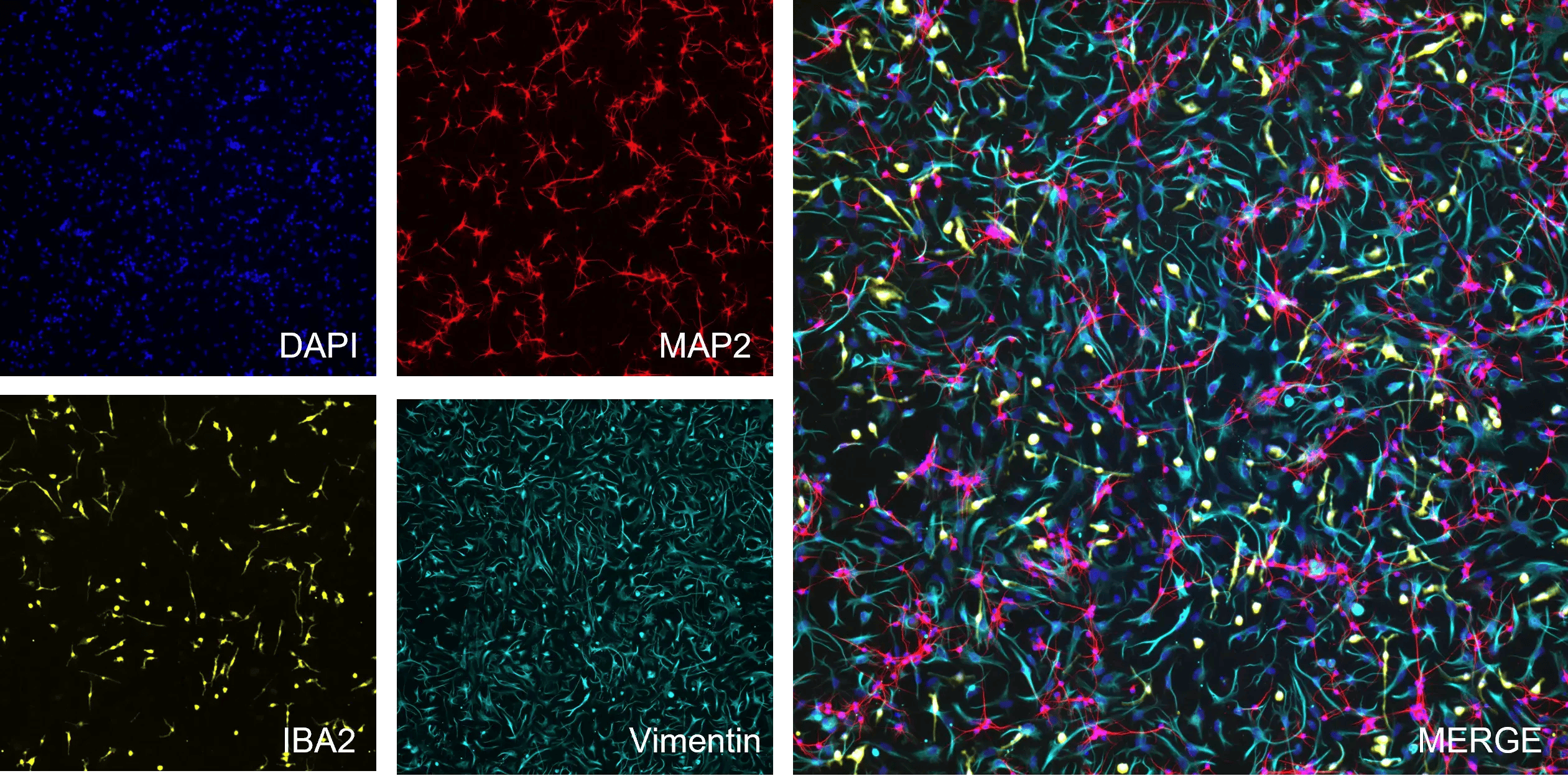
(UPDATED)ioAstrocytes_2.0_Hero_image_VIM_DAPI

Human iPSC-derived astrocytes

(UPDATED)ioAstrocytes_2.0_ioGlutamatergic-Neurons_co-culture_MEA-DIV50

Microelectrode array (MEA) functional characterisation of co-cultured ioGlutamatergic Neurons and ioAstrocytes 2.0

Representative MEA raster plots captured using the Axion BioSystems Maestro MEA platform evaluating co-cultures of 120K ioGlutamatergic Neurons and ioAstrocytes 2.0 (3:1 seeding ratio) over an extended maturation timeline (DIV29 to DIV50).

Each panel depicts a 5-minute continuous electrical activity recording. At early time points (DIV29–DIV34), cultures exhibit predominantly localised asynchronous neuronal firing. By DIV36–DIV41, co-cultures establish mature, highly synchronised functional networks, characterised by the emergence of organised network bursting (highlighted by orange vertical overlay markers).

Network synchrony is maintained through DIV50.

View the step-by-step ioGlutamatergic Neurons - ioAstrocytes 2.0 MEA protocol used to generate this data 

Phagocytosis of S. aureus by ioAstrocytes 2.0

Incucyte® video showing the ability of ioAstrocytes 2.0 to phagocytose pHrodo® Red S. aureus Bioparticles®.

pHrodo® Red S. aureus Bioparticles® were added to cultures of ioAstrocytes 2.0 at 7 days post-thaw. At the start of the video the particles are located outside the cells, due to the neutral pH of the media are non-fluorescent. But when phagocytosed, they are exposed to the acidic environments of intracellular organelles and fluoresce bright red.

The video shows an increase of red fluorescent particles accumulating within the cells over a time course of 48 hours, demonstrating that ioAstrocytes 2.0 have the capacity to phagocytose.

ioAstrocytes_2.0_cytokine-secretion_MSD_panel1

ioAstrocytes 2.0 secrete cytokines in response to stimulation

MSD multiplex immunoassay demonstrating that ioAstrocytes 2.0 secrete a range of cytokines upon treatment with various pro-inflammatory stimuli.

ioAstrocytes 2.0 were treated with 3 different pro-inflammatory cocktails and vehicle control at days 7 and 14 post-thaw. After 24 hours, media samples were collected to measure the concentration of cytokines in the media.

Data demonstrates that the pro-inflammatory cocktails induce secretion of most cytokines relative to the vehicle treated cells. Overall, ioAstrocytes 2.0 display the expected responses to the three distinct cocktails, including a strong response of Interleukin 6 (IL-6), known to be involved in neuroinflammation.

Note that IFNγ or IL-1β are present in two of the inflammatory cocktails and, therefore, the presence of these cytokines in the media will lead to higher signals upon their detection and interfere with the measurements of their secreted forms.

ioAstrocytes_2.0_cytokine-secretion_MSD_panel2

ioAstrocytes 2.0 secrete cytokines in response to stimulation

MSD multiplex immunoassay demonstrating that ioAstrocytes 2.0 secrete a range of cytokines upon treatment with various pro-inflammatory stimuli.

ioAstrocytes 2.0 were treated with 3 different pro-inflammatory cocktails and vehicle control at days 7 and 14 post-thaw. After 24 hours, media samples were collected to measure the concentration of cytokines in the media.

Data demonstrates that the pro-inflammatory cocktails induce secretion of most cytokines relative to the vehicle treated cells. Overall, ioAstrocytes 2.0 display the expected responses to the three distinct cocktails, including a strong response of Interleukin 6 (IL-6), known to be involved in neuroinflammation.

Note that IFNγ or IL-1β are present in two of the inflammatory cocktails and, therefore, the presence of these cytokines in the media will lead to higher signals upon their detection and interfere with the measurements of their secreted forms.

ioAstrocytes_2.0_ICC_Panel-1_DAPI_SOX9_S100B_D7_D14-1

Immunocytochemistry shows protein expression of key astrocyte markers

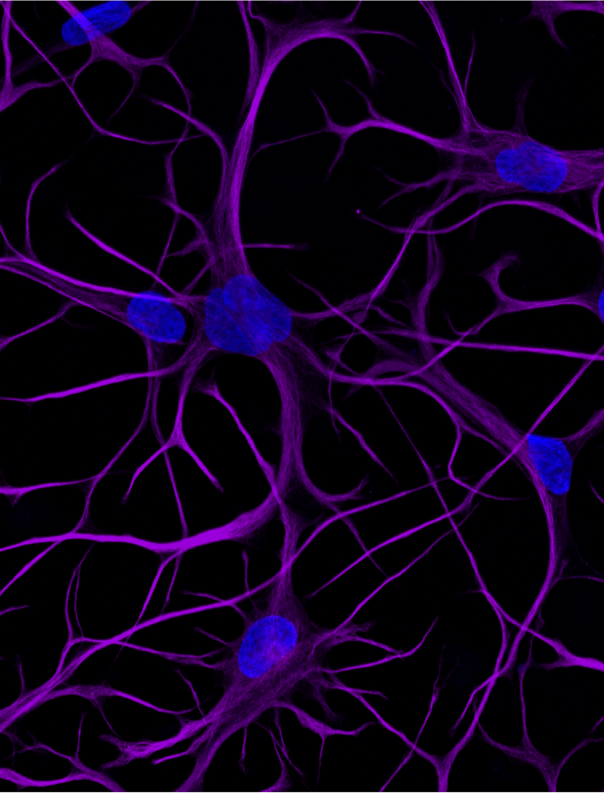
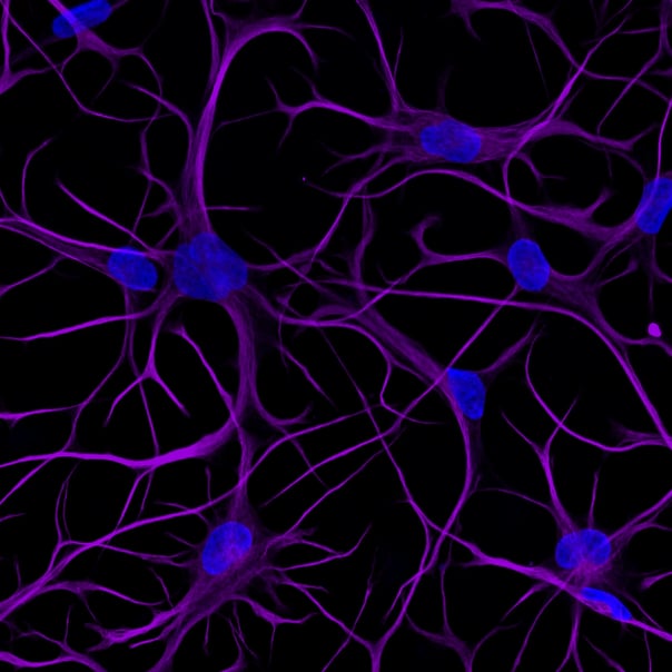
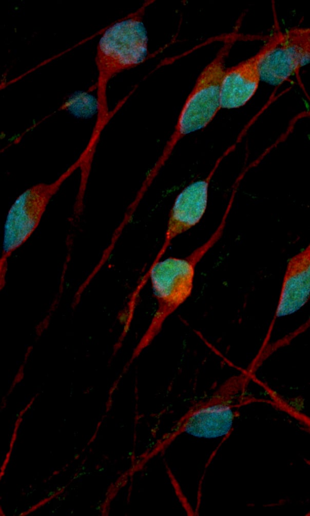
ioAstrocytes 2.0 express the key astrocyte markers S100B, SOX9 (A) and Vimentin (B) at days 7 and 14 post-thaw. DAPI was used as a nuclear stain

S100B is a multifaceted protein primarily found in astrocytes playing a key role in activation, neuroprotection, calcium homeostasis and astrocyte-neuron communication. SOX9 is critical for the differentiation of astrocytes.
ioAstrocytes_2.0_ICC_Panel-2_DAPI_VIM_MERGE_D7_D14-1

Immunocytochemistry shows protein expression of key astrocyte markers

ioAstrocytes 2.0 express the key astrocyte markers S100B, SOX9 (A) and Vimentin (B) at days 7 and 14 post-thaw. DAPI was used as a nuclear stain

S100B is a multifaceted protein primarily found in astrocytes playing a key role in activation, neuroprotection, calcium homeostasis and astrocyte-neuron communication. Vimentin is a cytoskeletal protein enriched in astrocytes. 

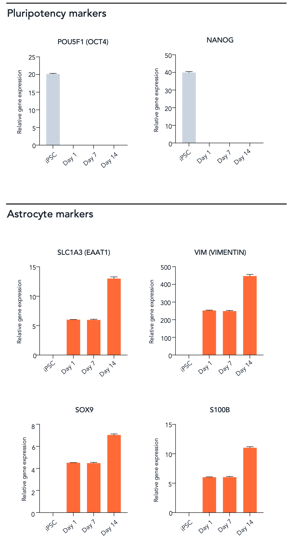
ioAstrocytes_2.0_RT-qPCR_NANOG_OCT4_EAAT1_VIM_SOX9_S100B_iPSC_D1_D7_D14

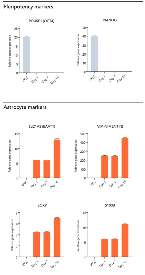
RT-qPCR shows gene expression of key astrocyte markers

RT-qPCR data showing expression of key astrocyte markers EAAT1, SOX9, S100B and Vimentin (VIM) at four different timepoints (iPSC & D1, D7, D14). ioAstrocytes 2.0 show expression of key markers from as early as day 1 post-thaw. Pluripotency markers POU5F1 (OCT4) & NANOG are downregulated.

The SLC1A3 gene encodes Excitatory Amino Acid Transporter 1 (EAAT1), a protein that plays a crucial role in regulating glutamate neurotransmission, maintaining neuronal health, and protecting against excitotoxicity.

View the step-by-step RNA extraction and RT-qPCR protocol used to generate this data

ioAstrocytes_ioGlutamatergic-Neurons_co-culture_DIV13

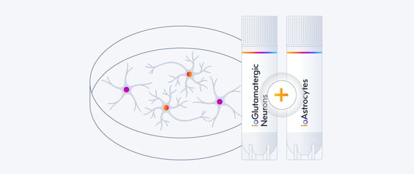
Easy-to-use co-culture protocol for ioAstrocytes 2.0 with ioGlutamatergic Neurons

The human brain is a complex and heterogeneous environment, containing multiple types of neurons and glial cells, each with a unique role to support the healthy function of the neuronal network. To make brain research as relevant as possible, it is important for researchers to create an in vitro environment that closely resembles the complex native in vivo environment, such as a co-culture of neuronal and glial cells. 

Access the protocol for co-culturing ioGlutamatergic Neurons and ioAstrocytes 2.0

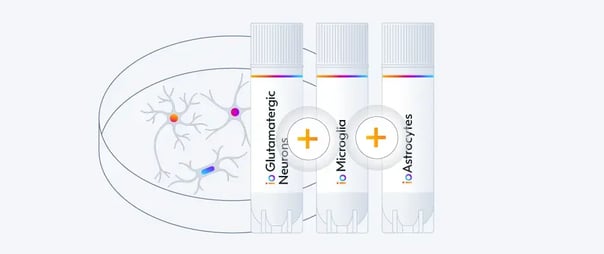
Phenotypic analysis of a tri-culture for CNS modelling using ioGlutamatergic Neurons, ioAstrocytes, and ioMicroglia

ioGlutamatergic Neurons, ioMicroglia and ioAstrocytes 2.0 tri-culture protocol

While co-cultures of neurons and astrocytes capture metabolic and structural synergy, the integration of microglia - the resident myeloid cells of the central nervous system (CNS), is critical for modelling neuroinflammatory pathways, synaptic pruning and homeostatic maintenance. 

Access the protocol for culturing ioMicroglia, ioGlutamatergic Neurons and ioAstrocytes 2.0

Vial limit exceeded

A maximum number of 20 vials applies. If you would like to order more than 20 vials, please contact us at orders@bit.bio.


Human iPSC-derived astrocytes

ioAstrocytes 2.0 are functional human iPSC-derived astrocytes, deterministically programmed using opti-ox technology. Delivered cryopreserved and ready for experiments in days, ioAstrocytes 2.0 adopt classic stellate morphology and express key markers including SOX9, EAAT1, S100B, and Vimentin.

Cells recapitulate critical human astrocytic functions - including phagocytosis, cytokine secretion, and MEA-validated modulation of neuronal activity. Maintained using serum-free culture components and supported by open-source protocols, ioAstrocytes 2.0 easily integrate into complex multi-cellular in vitro co-cultures alongside other CNS cell types, such as ioGlutamatergic Neurons and ioMicroglia.

ioAstrocytes 2.0 bridge translational gaps and overcome historical modelling limitations, delivering reproducible, human-relevant data to accelerate neurodegenerative disease research and therapeutic development.

Benchtop benefits

astrocytes_co-culture_ready

Co-culture ready

ioAstrocytes 2.0 support functional neuronal networks within co-culture settings, enabling in-vitro modelling of complex CNS biology.

astrocytes_functional_phagocytosis_cytokine_secretion

Functional

Display key phagocytic & cytokine secretion functions; alongside validated MEA functionality, demonstrating a direct influence on neuronal network activity.

astrocytes_consistency_data_reproducibility

Consistent

Get reproducible results from every vial with lot-to-lot consistency of highly characterised & defined human iPSC-derived cells.

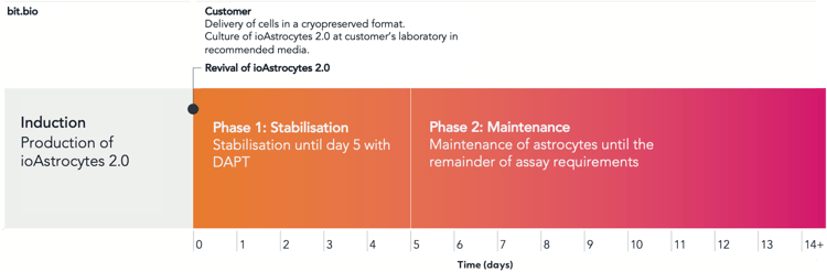
Schematic overview of the timeline in the user manual


ioAstrocytes_2.0_User_timelines

ioAstrocytes 2.0 are delivered in a cryopreserved format and are programmed to rapidly mature upon revival in the recommended serum-free media. The protocol for the generation of these cells is a two-phase process: 1. Stabilisation for 5 days. 2. From day 5 onwards, maintenance of cells according to the protocol and recommended media for the duration of assay requirements.

Product specifications

Starting material

Human iPSC line

Seeding compatibility

6, 12, 24 and 96 well plates

Shipping info

Dry ice

Donor

Caucasian adult male (skin fibroblast),
Genotype APOE 3/4

Vial size

Small: ≥2 x 10⁶ viable cells

Quality control

Sterility, protein expression (ICC) and gene expression (RT-qPCR)

Differentiation method

opti-ox deterministic cell programming

Recommended seeding density

45,000 cells/cm²

User storage

LN2 or -150°C

Format

Cryopreserved cells

Product use

ioCells are for research use only

Applications

Neurodegenerative disease modelling
Drug screening & development
Neuropharmacology
Neuroinflammation research
Biomarker discovery
Neurotoxicology


Scale your study with volume pricing

Enabling scientists to use human cells in their research, running additional experiments without rationing cells or limiting experimental scale


Order quantity Total vials received Pricing tier 
1 - 9 packs 2 - 18 vials Standard price
10 - 49 packs 20 - 98 vials Automatic 10% discount
> 50 packs > 100 vials > Contact us for a quote


Academic pricing: Academic users can purchase any ioCells in 3-vial packs ($/€/£ 999 per pack), available year-round with any cell type combination.

What scientists say about ioAstrocytes

An image of Jeremy Krohn

Jeremy Krohn

PhD Candidate | DZNE / Charité University of Medicine

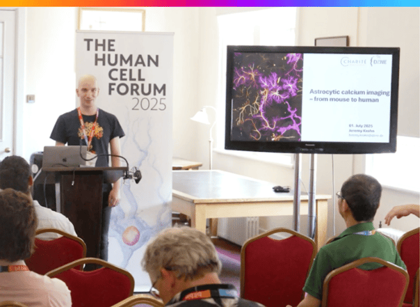
"We used bit.bio human ioAstrocytes in our experiments to compare with - and step-wise replace - animal derived cells. The human cells were straightforward to establish, showed high reproducibility, and performed well in calcium imaging experiment."

Technical data

Rapid gain of functional activity

ioAstrocytes 2.0 support neuronal co-cultures and enhance MEA activity

(UPDATED)ioAstrocytes_2.0_ioGlutamatergic-Neurons_co-culture_MEA-DIV50

Representative MEA raster plots captured using the Axion BioSystems Maestro MEA platform evaluating co-cultures of 120K ioGlutamatergic Neurons and ioAstrocytes 2.0 (3:1 seeding ratio) over an extended maturation timeline (DIV29 to DIV50).

Each panel depicts a 5-minute continuous electrical activity recording. At early time points (DIV29–DIV34), cultures exhibit predominantly localised asynchronous neuronal firing. By DIV36–DIV41, co-cultures establish mature, highly synchronised functional networks, characterised by the emergence of organised network bursting (highlighted by orange vertical overlay markers). Network synchrony is maintained through DIV50.

View the step-by-step ioGlutamatergic Neurons - ioAstrocytes 2.0 MEA protocol used to generate this data

Phagocytosis of S. aureus by ioAstrocytes 2.0

Incucyte® video showing the ability of ioAstrocytes 2.0 to phagocytose pHrodo® Red S. aureus Bioparticles®.

pHrodo® Red S. aureus Bioparticles® were added to cultures of ioAstrocytes 2.0 at 7 days post-thaw. At the start of the video the particles are located outside the cells, due to the neutral pH of the media are non-fluorescent. But when phagocytosed, they are exposed to the acidic environments of intracellular organelles and fluoresce bright red.

The video shows an increase of red fluorescent particles accumulating within the cells over a time course of 48 hours, demonstrating that ioAstrocytes 2.0 have the capacity to phagocytose.

ioAstrocytes 2.0 secrete cytokines in response to stimulation
ioAstrocytes_2.0_cytokine-secretion_MSD_panel1
ioAstrocytes_2.0_cytokine-secretion_MSD_panel2

Click on the tabs to explore the data.

MSD multiplex immunoassay demonstrating that ioAstrocytes 2.0 secrete a range of cytokines upon treatment with various pro-inflammatory stimuli.

ioAstrocytes 2.0 were treated with 3 different pro-inflammatory cocktails and vehicle control at days 7 and 14 post-thaw. After 24 hours, media samples were collected to measure the concentration of cytokines in the media.

Data demonstrates that the pro-inflammatory cocktails induce secretion of most cytokines relative to the vehicle treated cells. Overall, ioAstrocytes 2.0 display the expected responses to the three distinct cocktails, including a strong response of Interleukin 6 (IL-6), known to be involved in neuroinflammation. 

Note that IFNγ or IL-1β are present in two of the inflammatory cocktails and, therefore, the presence of these cytokines in the media will lead to higher signals upon their detection and intefere with the measurements of their secreted forms.

Highly characterised and defined

Immunocytochemistry shows protein expression of key astrocyte markers

ioAstrocytes_2.0_ICC_Panel-1_DAPI_SOX9_S100B_D7_D14-1
ioAstrocytes_2.0_ICC_Panel-2_DAPI_VIM_MERGE_D7_D14-1

Click on the tabs to explore the data.

ioAstrocytes 2.0 express the key astrocyte markers S100B, SOX9 (A) and Vimentin (B) at days 7 and 14 post-thaw.  DAPI was used as a nuclear stain

S100B is a multifaceted protein primarily found in astrocytes playing a key role in activation, neuroprotection, calcium homeostasis and astrocyte-neuron communication. SOX9 is critical for the differentiation of astrocytes. Vimentin is a cytoskeletal protein enriched in astrocytes.

RT-qPCR shows gene expression of key astrocyte markers
ioAstrocytes_2.0_RT-qPCR_NANOG_OCT4_EAAT1_VIM_SOX9_S100B_iPSC_D1_D7_D14

RT-qPCR data showing expression of key astrocyte markers EAAT1, SOX9, S100B and Vimentin (VIM) at four different timepoints (iPSC & D1, D7, D14). ioAstrocytes 2.0 show expression of key markers from as early as day 1 post-thaw. Pluripotency markers POU5F1 (OCT4) & NANOG are downregulated.

The SLC1A3 gene encodes Excitatory Amino Acid Transporter 1 (EAAT1), a protein that plays a crucial role in regulating glutamate neurotransmission, maintaining neuronal health, and protecting against excitotoxicity.

View the step-by-step RNA extraction and RT-qPCR protocol used to generate this data  

Technical data

Functional MEA co-culture

MEA co-culture protocol for ioGlutamatergic Neurons and ioAstrocytes 2.0
(UPDATED)ioAstrocytes_2.0_ioGlutamatergic-Neurons_co-culture_MEA-DIV50

ioGlutamatergic Neuron–ioAstrocyte 2.0 co-cultures form mature, synchronised neuronal networks over time.

Representative MEA raster plots generated using the Axion BioSystems Maestro MEA platform show electrical activity in co-cultures of 120K ioGlutamatergic Neurons and ioAstrocytes 2.0, seeded at a 3:1 ratio, across an extended maturation period (DIV29–DIV50). Each panel represents a 5-minute continuous recording.

At early time points (DIV29–DIV34), cultures display predominantly localised, asynchronous neuronal firing. From DIV36–DIV41, co-cultures develop mature, highly synchronised functional networks, characterised by organised network bursting (orange vertical markers), with network synchrony maintained through DIV50.

In-vitro CNS co-culture model

Easy-to-use co-culture protocol for ioAstrocytes 2.0 with ioGlutamatergic Neurons 
ioAstrocytes_ioGlutamatergic-Neurons_co-culture_DIV13

The human brain is a complex and heterogeneous environment, containing multiple types of neurons and glial cells, each with a unique role to support the healthy function of the neuronal network. To make brain research as relevant as possible, it is important for researchers to create an in vitro environment that closely resembles the complex native in vivo environment, such as a co-culture of neuronal and glial cells.

In vitro CNS tri-culture model

ioGlutamatergic Neurons, ioMicroglia and ioAstrocytes 2.0 tri-culture protocol

Phenotypic analysis of a tri-culture for CNS modelling using ioGlutamatergic Neurons, ioAstrocytes, and ioMicroglia

While co-cultures of neurons and astrocytes capture metabolic and structural synergy, the integration of microglia - the resident myeloid cells of the central nervous system (CNS), is critical for modelling neuroinflammatory pathways, synaptic pruning and homeostatic maintenance.

Frequently Asked Questions (FAQs)

What is the different between ioAstrocytes and ioAstrocytes 2.0?

ioAstrocytes 2.0 builds on the original ioAstrocytes product with an optimised workflow, expanded functional characterisation and enhanced support for advanced CNS applications, including MEA. With demonstrated utility in neuronal co-culture, functional network and neuroinflammation studies, ioAstrocytes 2.0 provides a robust and reproducible human astrocyte model for building more physiologically relevant in vitro systems.


How long can ioAstrocytes 2.0 be kept in culture?

ioAstrocytes 2.0 have been maintained up to at least 2 weeks in monoculture following culture conditions recommended in the User Manual without impairment to function and culture attachment.


Can ioAstrocytes 2.0 be utilised to study neuroglial interactions?

ioAstrocytes can be cultured with other glial cells and neurons to study neuroglial interactions. bit.bio has optimised protocols for co-culturing ioAstrocytes 2.0 with ioGlutamatergic Neurons and a tri-culture protocol for the culture of ioAstrocytes 2.0 with ioGlutamatergic Neurons.


Can ioAstrocytes 2.0 be used for functional assays, such as MEA?

ioAstrocytes 2.0 support neuronal co-cultures and establish mature, highly synchronous functional networks. bit.bio has a step-by-step validated MEA protocol for the co-culture of our ioAstrocytes 2.0 with ioGlutamatergic Neurons.


Expand your research

Click on the icons to find out more

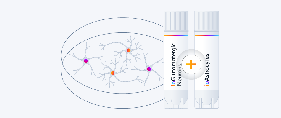
Co-culture hiPSC-derived glutamatergic neurons and astrocytes
Build neurodegeneration disease models
Model neurodegeneration in vitro with astrocyte-neuron co-cultures
Expand your research
Build neurodegeneration disease models
Model neurodegeneration in vitro with astrocyte-neuron co-cultures
Co-culture hiPSC-derived glutamatergic neurons and astrocytes

Access 20 neuronal disease models with a single co-culture protocol.

View the co-culture protocol
Explore ioGlutamatergic Neuron Disease Models 

Multi-cellular model with neurons and glia human iPSC-derived cells.
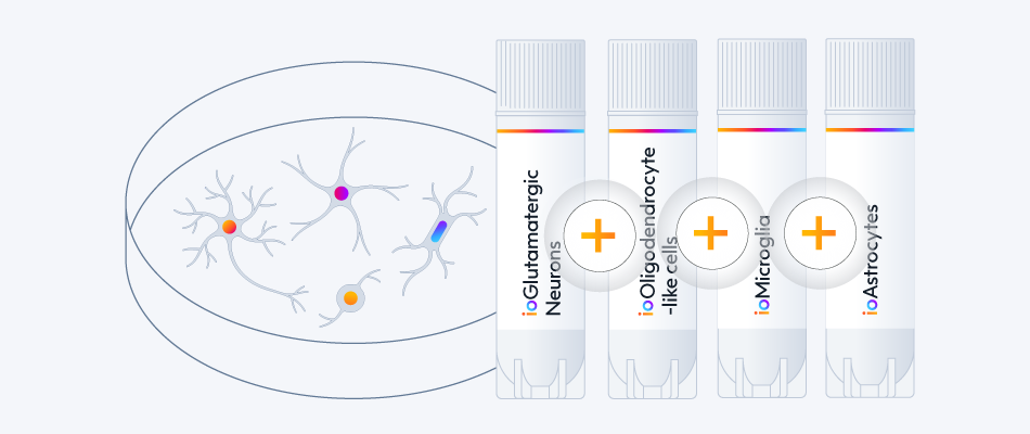
Generate functional in vitro models of the CNS
Highly pure human iPSC-derived cells to build complex multi-cell cultures
Expand your research
Generate functional in vitro models of the CNS
Highly pure human iPSC-derived cells to build complex multi-cell cultures
Multi-cellular model with neurons and glia human iPSC-derived cells.

ioAstrocytes are highly pure, defined and consistent, a great companion for your CNS model. 
Combine them with other neuronal and glia ioCells to generate complex multi-cell cultures. 

ioGlutamatergic Neurons 
ioMicroglia
ioOligodendrocyte-like cells

Multi-cellular model with neurons and glia human iPSC-derived cells.
Study neuroinflammation
Assess cell damage, microglia activation and cytokine release
Expand your research
Study neuroinflammation
Assess cell damage, microglia activation and cytokine release
Multi-cellular model with neurons and glia human iPSC-derived cells.

Interested in optimising your neuroinflammation studies?
Move into a multi-cellular model using different cell types:

ioMicroglia Male
ioGlutamatergic Neurons
ioOligodendrocyte-like cells

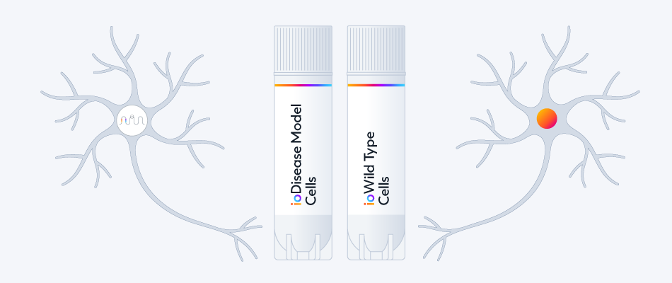
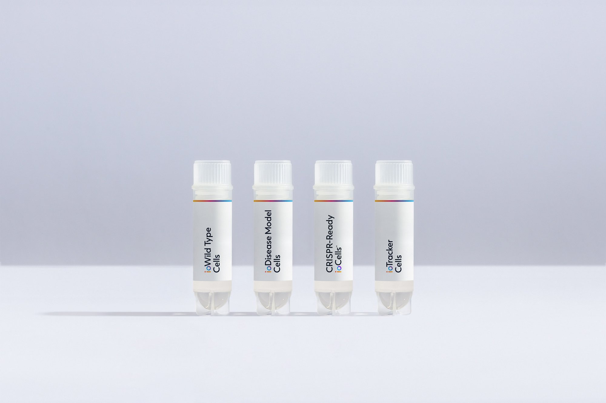
Tailor experimental conditions to answer your scientific question by including ioDisease Model Cells with disease-related mutations.

Modeling disease with hiPSC-derived astrocytes, comparing disease-related mutations with an isogenic control.
Study disease-related mutations
Make true comparisons with a genetically-matched control
Expand your research
Study disease-related mutations
Make true comparisons with a genetically-matched control
Modeling disease with hiPSC-derived astrocytes, comparing disease-related mutations with an isogenic control.

Build your custom disease model into the ioAstrocytes wild type. Use the genetically matched control in your experiments to make true comparisons in your data, being confident that even subtle variances in your data are attributable to your mutation of interest.

Start the conversation today

ioCells catalogue

Human iPSC-derived cells

powered by opti-ox

Consistent. Defined. Scalable.

bitbio-cell_catalogue_header-with-tracker-Desktop-2500x1664