cat no | 02-3002-0000
Cryopreserved human iPSC-derived cardiomyocytes with a TTN A-band mutation, generated by directed differentiation, and ready for experiments in days
Human iPSC-derived ventricular cardiomyocytes carrying a TTN A-band frameshift mutation
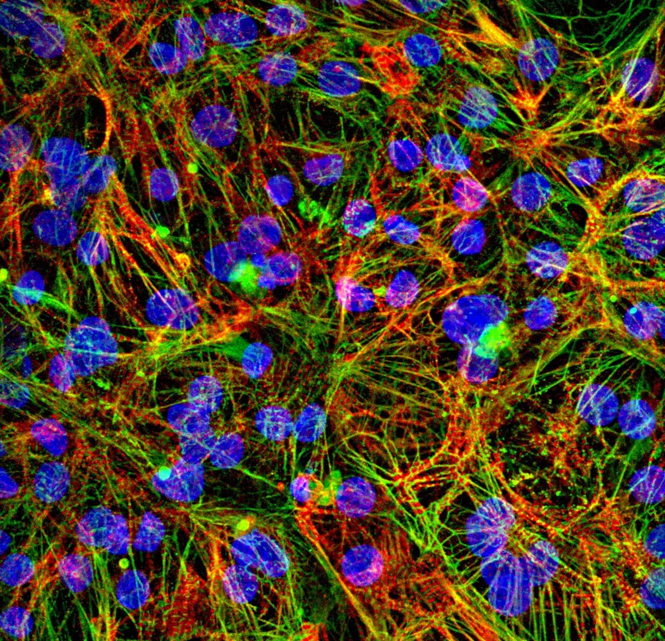
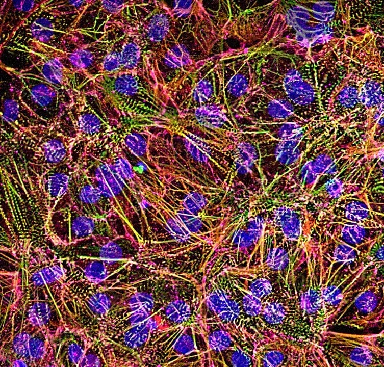
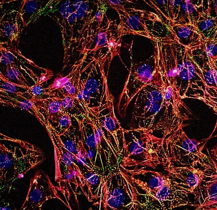
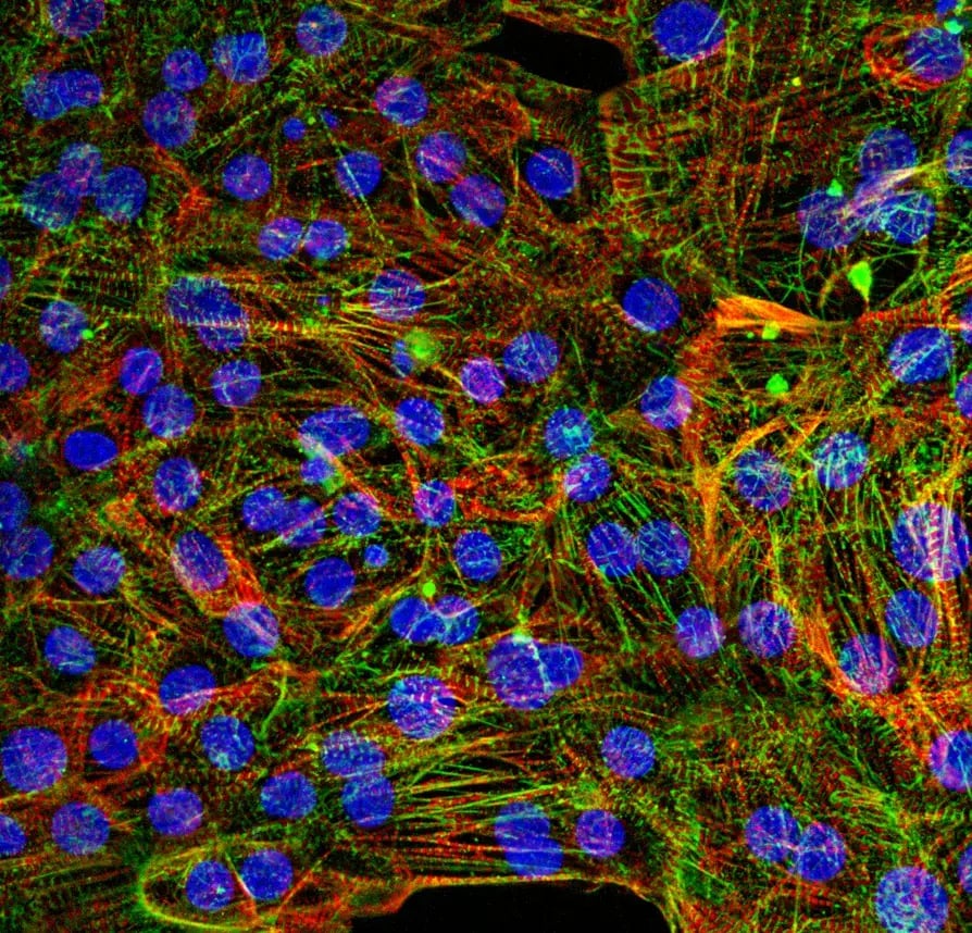
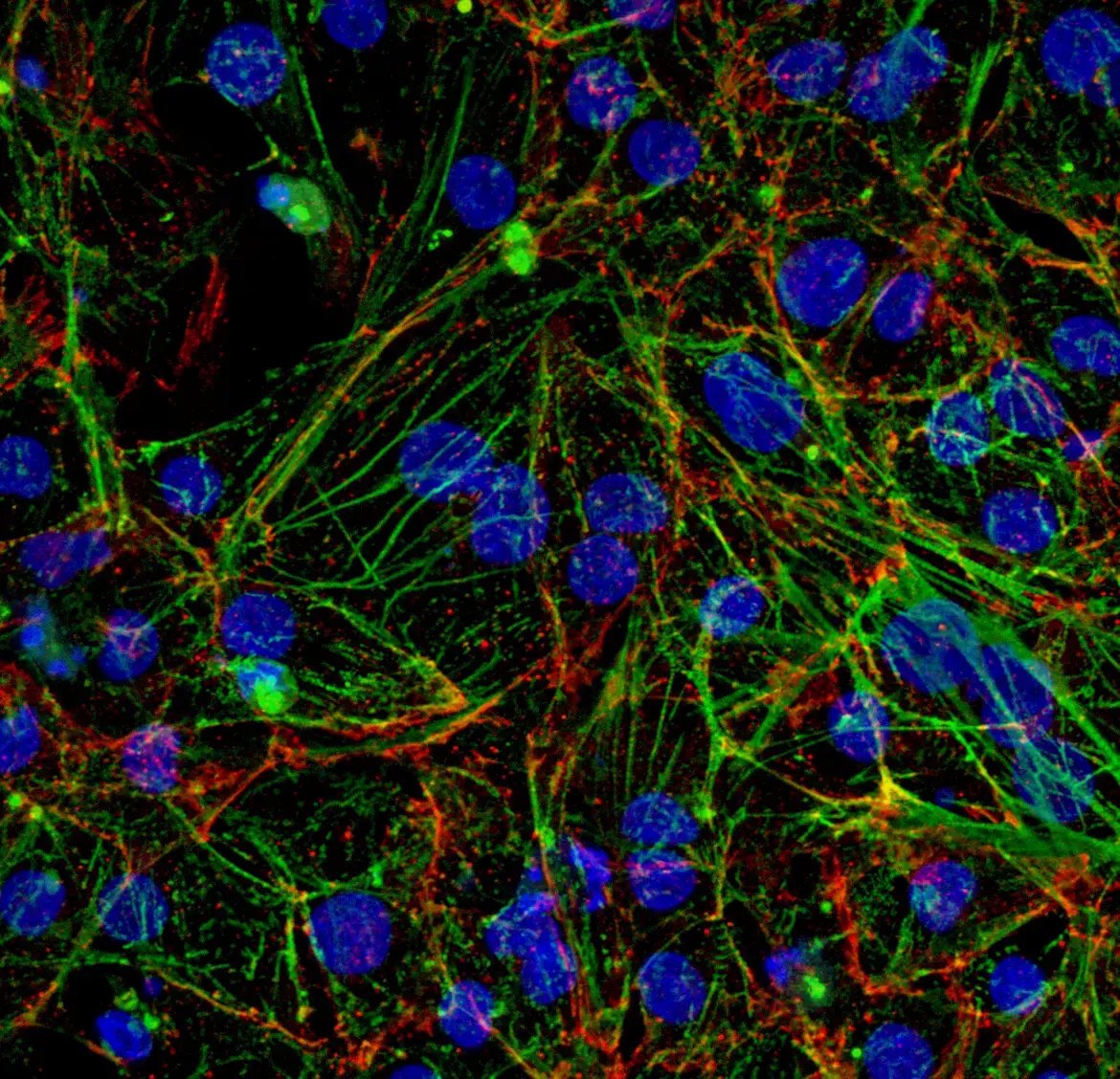
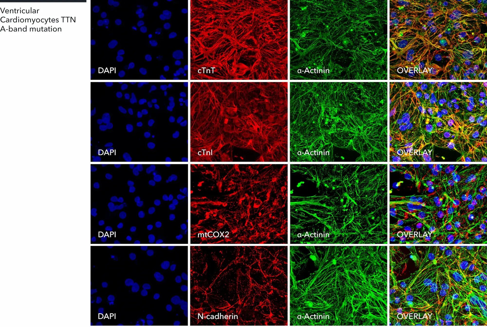
Ventricular Cardiomyocytes TTN A-band mutation express key cardiac specific markers
Immunofluorescent staining confirms cardiomyocyte identity by robust expression of α-actinin, cardiac troponin T (cTnT), and cardiac troponin I (cTnl), with additional staining for mitochondria (mtCOX2) and N-cadherin. Notably, α-actinin staining reveals disrupted sarcomeric organisation and altered sarcomere alignment in the TTN A-band mutant cardiomyocytes, demonstrating a disease-associated structural phenotype. Nuclei are counterstained with DAPI; 60x magnification.
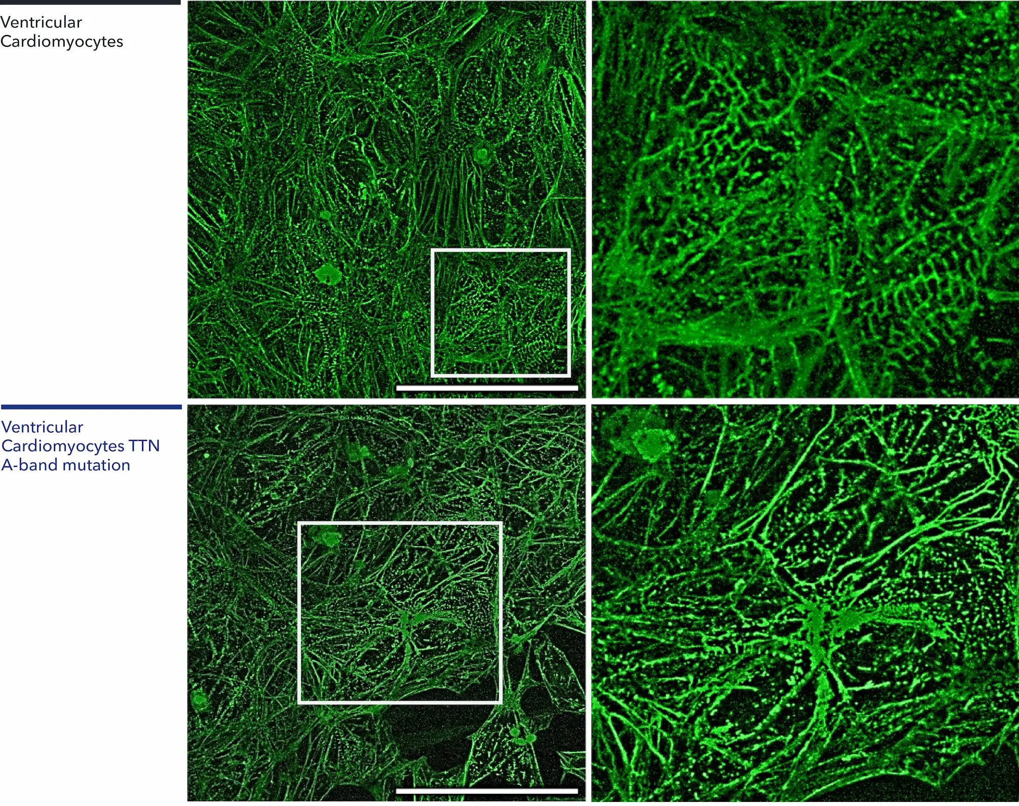
Distinctive sarcomeric disorganisation in Ventricular Cardiomyocytes TTN A-band mutation
Immunofluorescent staining of α-actinin at 10 days post-thaw reveals robust expression in both wild-type and TTN A-band mutant Ventricular Cardiomyocytes. However, compared to the well-organised striations in the wild-type control (top row), the TTN A-band mutant cardiomyocytes (bottom row) display a disorganised sarcomeric architecture. White boxes indicate regions magnified in the right-hand panels to detail these structural differences. Scale bar: 50 μm, 40x magnification.
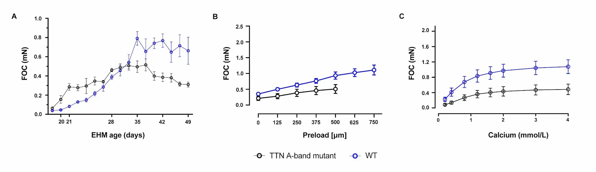
TTN A-band mutation impairs longitudinal contractility in 3D engineered heart muscle
Human iPSC-derived cardiomyocytes were combined with fibroblasts in a collagen hydrogel to generate wild-type (WT) and TTN A-band heterozygous mutant (TTN A-band mutant) engineered heart muscle (EHM). Tissue function was assessed over 48 days of maturation via live longitudinal video-optical tracking (myrImager), utilising automated peak finding and pole detection. The EHMs were subsequently analysed in an organ bath under defined isometric conditions for deep functional validation at the study endpoint.
Force of contraction (FOC) measurements demonstrate a progressive improvement in WT tissues over time, whereas TTN A-band mutant EHMs display a declining trend in FOC, indicating impaired contractile function (A).
In the endpoint organ bath analysis, WT EHMs demonstrate a progressive increase in force with increasing preload, consistent with a functional Frank–Starling response, whereas TTN A-band mutant EHMs show markedly reduced preload-dependent force generation (B).
Deep phenotyping of contractility at day 48 demonstrates a reduced Ca²⁺-dependent force response in TTN A-band mutant EHMs compared with WT controls, indicating impaired contractile reserve (C).
Data shown as mean ± SEM, n=8.
A maximum number of 20 vials applies. If you would like to order more than 20 vials, please contact us at orders@bit.bio.

myrCell Ventricular Cardiomyocytes TTN mutation are human iPSC-derived cells generated using traditional differentiation protocols. The cells carry a heterozygous frameshift mutation in the TTN A-band (c.70692_70693insAT/WT), which is widely recognised as a gold-standard, highly penetrant variant for modelling dilated cardiomyopathy (DCM).
This disease model is suitable for generation of 3D engineered heart muscle and demonstrates reduced contractility compared to the wild-type control, making it an ideal platform for investigating disease mechanisms and screening novel cardioprotective therapeutics.
The cells can be paired with a genetically matched wild-type control and are also available with an integrated live sarcomere reporter to facilitate advanced structural analysis.
Make true comparisons
Pair the DCM disease model cells with the wild-type control to investigate the impact of the TTN A-band mutation.
Functional
Cells contract in 2D and 3D, beat spontaneously, exhibit key cardiac markers and ventricular-specific electrophysiology.
Quick
Ready to use for functional experiments within 7 days post-revival.
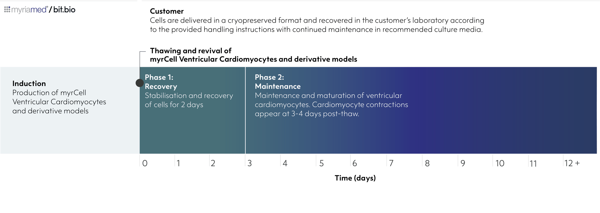
Ventricular Cardiomyocytes TTN A-band mutation are delivered in a cryopreserved format and recovered in the customer’s laboratory according to the provided handling instructions with continued maintenance in recommended culture media.
Starting material
Human iPSC line, W001
Karyotype
Normal (46, XY)
Seeding compatibility
6, 12, 24, 48,96 & 384 well plates
Shipping info
Dry ice
Donor
Male
Vial size
Small: >1 x 10⁶ viable cells
Quality control
Sterility, protein expression (ICC)
Differentiation method
Small molecule-directed differentiation
Recommended minimum seeding density
90,000 cells/cm² (laminin-coated plates); 150,000 cells/cm² (Matrigel-coated plates)
User storage
LN2 or -150°C
Format
Cryopreserved cells
Genetic modification
Heterozygous TTN A-band frameshift mutation (c.70692_70693insAT/WT)
Applications
Cardiovascular research
Disease modelling
2D screening and functional assays
3D engineered heart muscle
Product use
myrCells are for research use only
Enabling scientists to use human cells in their research, running additional experiments without rationing cells or limiting experimental scale
| Order quantity | Total vials received | Pricing tier |
| 1 - 9 packs | 3 - 27 vials | Standard price |
| 10 - 33 packs | 30 - 99 vials | Automatic 10% discount |
| > 34 packs | > 100 vials | > Contact us for a quote |
Ventricular Cardiomyocytes TTN A-band mutation express key cardiac specific markers
Immunofluorescent staining confirms cardiomyocyte identity by robust expression of α-actinin, cardiac troponin T (cTnT), and cardiac troponin I (cTnl), with additional staining for mitochondria (mtCOX2) and N-cadherin. Notably, α-actinin staining reveals disrupted sarcomeric organisation and altered sarcomere alignment in the TTN A-band mutant cardiomyocytes, demonstrating a disease-associated structural phenotype. Nuclei are counterstained with DAPI; 60x magnification.
Distinctive sarcomeric disorganisation in Ventricular Cardiomyocytes TTN A-band mutation
Immunofluorescent staining of α-actinin at 10 days post-thaw reveals robust expression in both wild-type and TTN A-band mutant Ventricular Cardiomyocytes. However, compared to the well-organised striations in the wild-type control (top row), the TTN A-band mutant cardiomyocytes (bottom row) display a disorganised sarcomeric architecture. White boxes indicate regions magnified in the right-hand panels to detail these structural differences. Scale bar: 50 μm, 40x magnification.
TTN A-band mutation impairs longitudinal contractility in 3D engineered heart muscle
Human iPSC-derived cardiomyocytes were combined with fibroblasts in a collagen hydrogel to generate wild-type (WT) and TTN A-band heterozygous mutant (TTN A-band mutant) engineered heart muscle (EHM). Tissue function was assessed over 48 days of maturation via live longitudinal video-optical tracking (myrImager), utilising automated peak finding and pole detection. The EHMs were subsequently analysed in an organ bath under defined isometric conditions for deep functional validation at the study endpoint.
Force of contraction (FOC) measurements demonstrate a progressive improvement in WT tissues over time, whereas TTN A-band mutant EHMs display a declining trend in FOC, indicating impaired contractile function (A).
In the endpoint organ bath analysis, WT EHMs demonstrate a progressive increase in force with increasing preload, consistent with a functional Frank–Starling response, whereas TTN A-band mutant EHMs show markedly reduced preload-dependent force generation (B).
Deep phenotyping of contractility at day 48 demonstrates a reduced Ca²⁺-dependent force response in TTN A-band mutant EHMs compared with WT controls, indicating impaired contractile reserve (C).
Data shown as mean ± SEM, n=8.
The A-band frameshift mutation (c.70692_70693insAT) in the TTN gene is widely recognised as a prototypical, highly penetrant variant for modelling genetic Dilated Cardiomyopathy (DCM). Occurring in the constitutively expressed A-band of the titin protein, this mutation reliably triggers the two primary drivers of heart failure in patients: a lack of functional titin to support contraction (haploinsufficiency) and severe cellular stress from a toxic buildup of truncated proteins (proteotoxicity). Human iPSC-derived cardiomyocytes carrying this specific mutation provide a reliable platform for recreating the molecular and functional hallmarks of heart failure in vitro.
The Ventricular Cardiomyocytes TTN A-band mutation disease model is offered alongside a genetically-matched, wild-type control. Together, these paired cells provide a controlled, physiologically relevant model for investigating the impact of the frameshift mutation on cellular and molecular mechanisms and function.
The Ventricular Cardiomyocytes TTN A-band mutation model is designed for dilated cardiomyopathy (DCM) research. It is ideal for disease phenotyping, exploring sarcomere biology, target validation, and therapeutic screening. The cells are highly adaptable, with proven applications spanning both 2D cellular assays and 3D engineered heart muscle (EHM) models.
Consistent. Defined. Scalable.