myrCell SarcPaint Ventricular Cardiomyocytes
myrcell-ventricular-cardiomyocyte-sarcpaint-alpha-actinin-n-cadherin
myrcell-ventricular-cardiomyocyte-sarcpaint-live-imaging-60x
myrcell-ventricular-cardiomyocytes-z-band-profiles
myrcell-sarcpaint-ventricular-cardiomyocytes-icc-ctnt-ctni-mlc2v
myrcell-sarcpaint-ventricular-cardiomyocytes-icc-mtcox2-n-cadherin-cx43
myrcell-ventricular-cardiomyocyte-sarcpaint-sarcomere-remodelling
myrcell-ventricular-cardiomyocyte-sarcpaint-drug-induced-sarcomere-remodelling-video-1x4
myrcell-ventricular-cardiomyocytes-sarcpaint-compound-screen
myrcell-ventricular-cardiomyocytes-sarcpaint-phenotypic-screening
myrcell-cardiomyocytes-ehm-contraction-thumbnail
myrcell-ventricular-cardiomyocytes-sarcpaint-ehm-protein-expression
myrcell-ventricular-cardiomyocytes-sarcpaint-ehm-video-optics-organ-bath
myrcell-ventricular-cardiomyocyte-ehm-contractility-wt-sarcpaint
myrcell-ventricular-cardiomyocyte-sarcpaint-alpha-actinin-n-cadherin
myrcell-ventricular-cardiomyocyte-sarcpaint-live-imaging-60x
myrcell-ventricular-cardiomyocytes-z-band-profiles
myrcell-sarcpaint-ventricular-cardiomyocytes-icc-ctnt-ctni-mlc2v
myrcell-sarcpaint-ventricular-cardiomyocytes-icc-mtcox2-n-cadherin-cx43
myrcell-ventricular-cardiomyocyte-sarcpaint-sarcomere-remodelling
myrcell-ventricular-cardiomyocyte-sarcpaint-drug-induced-sarcomere-remodelling-video-1x4
myrcell-ventricular-cardiomyocytes-sarcpaint-compound-screen
myrcell-ventricular-cardiomyocytes-sarcpaint-phenotypic-screening
myrcell-cardiomyocytes-ehm-contraction-thumbnail
myrcell-ventricular-cardiomyocytes-sarcpaint-ehm-protein-expression
myrcell-ventricular-cardiomyocytes-sarcpaint-ehm-video-optics-organ-bath
myrcell-ventricular-cardiomyocyte-ehm-contractility-wt-sarcpaint

cat no | 02-5001-0000

myrCell SarcPaint Ventricular Cardiomyocytes

Human iPSC-derived ventricular cardiomyocytes with a live sarcomere reporter

  • Cryopreserved human iPSC-derived ventricular cardiomyocytes stably expressing a genetically encoded ACTN2-Citrine reporter, generated by directed differentiation, and ready for experiments in days

  • Enables live, label-free imaging and quantification of sarcomere organisation; ideal for longitudinal high-content imaging and high-throughput screening

  • Highly characterised to show functional synchronised beating by day 7 post-revival
myrcell-ventricular-cardiomyocyte-sarcpaint-alpha-actinin-n-cadherin

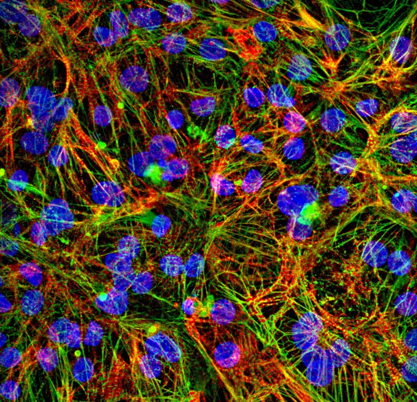
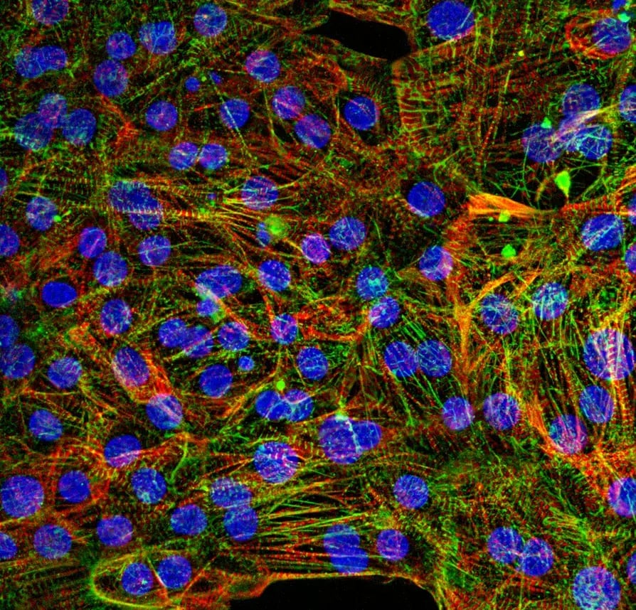
Human iPSC-derived ventricular cardiomyocytes with SarcPaint reporter

Immunofluorescence staining of human iPSC-derived SarcPaint Ventricular Cardiomyocytes 10 days post-thawing showing α-actinin (endogenous Citrine reporter; green), N-cadherin (magenta), and α-actinin immunostaining (red), with nuclei counterstained with DAPI (blue). Alpha-actinin is a key structural component of the sarcomere and its organised localisation reflects the development of the contractile apparatus. N-cadherin is an important component of cardiac adherens junctions, mediating cell–cell adhesion and contributing to the structural and functional coupling of cardiomyocytes. The co-localisation of the endogenous α-actinin-Citrine reporter and α-actinin immunostaining appears yellow in the merged channels, confirming the specificity of the reporter signal. 60x magnification.

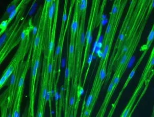
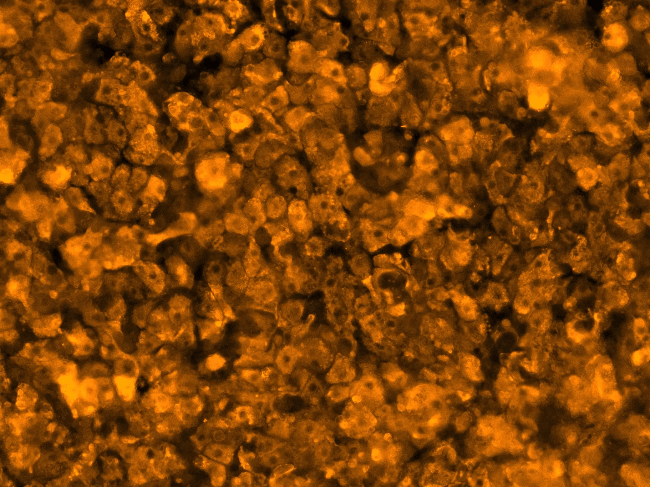
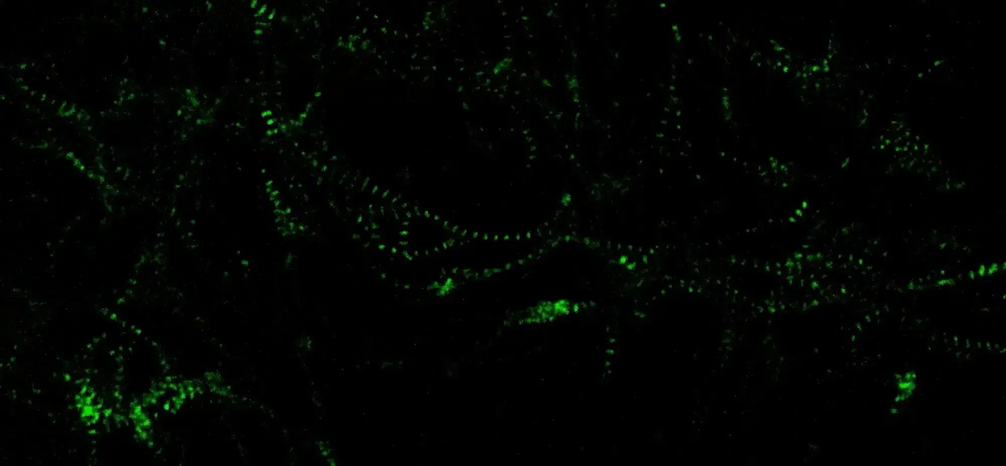
Real-time imaging of sarcomeric dynamics in SarcPaint Ventricular Cardiomyocytes

Live fluorescent recording of SarcPaint Ventricular Cardiomyocytes, cultured in Matrigel-coated flasks; recorded at Day 10 post-thaw. Scale: 100 μm, 60x magnification. 

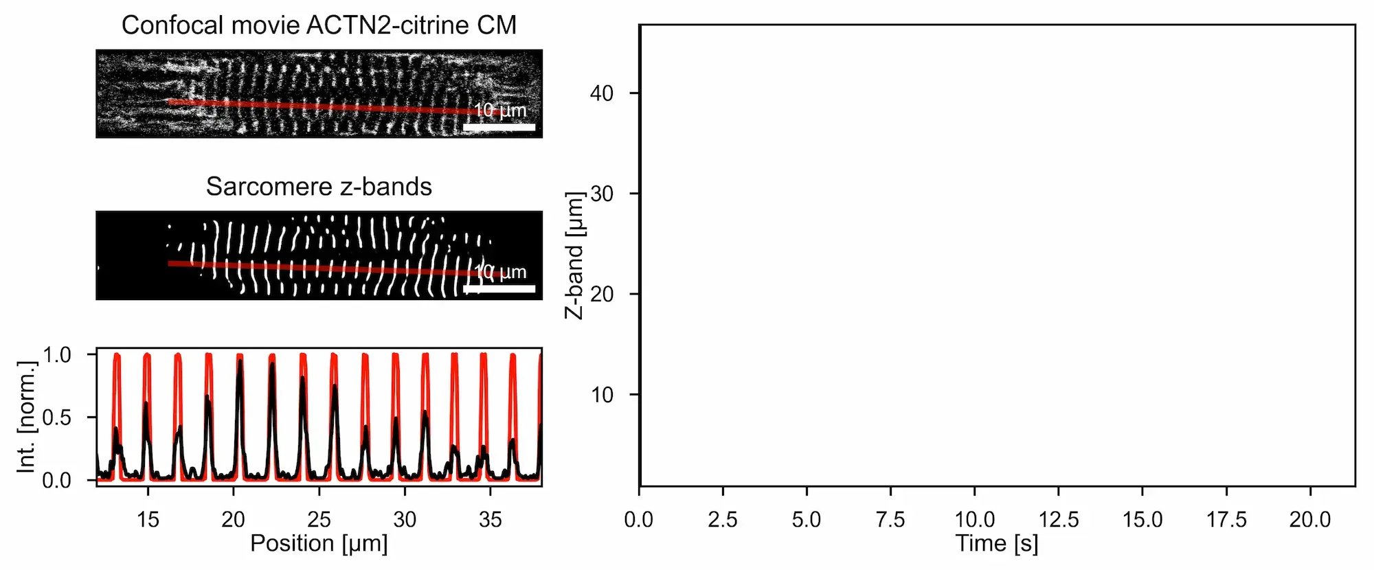
Real-time monitoring and longitudinal analysis of sarcomere organisation and function

Automated extraction of Z-band profiles and quantitative measurement of sarcomere spacing from confocal images enabling deep learning-based segmentation and spatial mapping with automated AI assisted workflows. This model enables automated multi-scale quantification of sarcomere organisation, myofibril architecture, and contractile dynamics directly from live-cell images and videos, thus eliminating endpoint staining while supporting longitudinal phenotyping and high-content drug screening. 

myrcell-sarcpaint-ventricular-cardiomyocytes-icc-ctnt-ctni-mlc2v

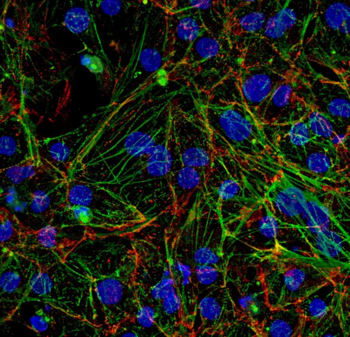
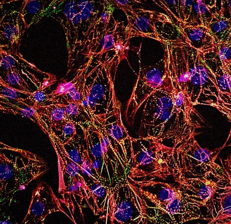
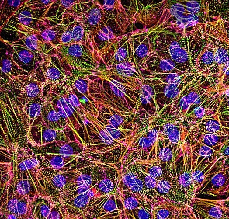
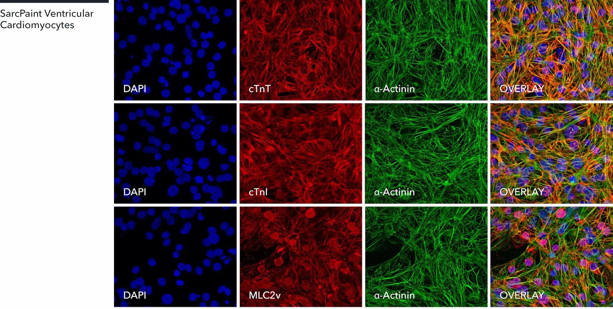
SarcPaint Ventricular Cardiomyocytes express key cardiac specific markers

Immunofluorescent staining of human iPSC-derived SarcPaint Ventricular Cardiomyocytes at 10 days post-thawing showing SarcPaint-tagged α-actinin (green) with co-localisation of cardiac troponin T (cTnT), cardiac troponin I (cTnI) or ventricular myosin light chain-2 (MLC2v); 60x magnification.

myrcell-sarcpaint-ventricular-cardiomyocytes-icc-mtcox2-n-cadherin-cx43

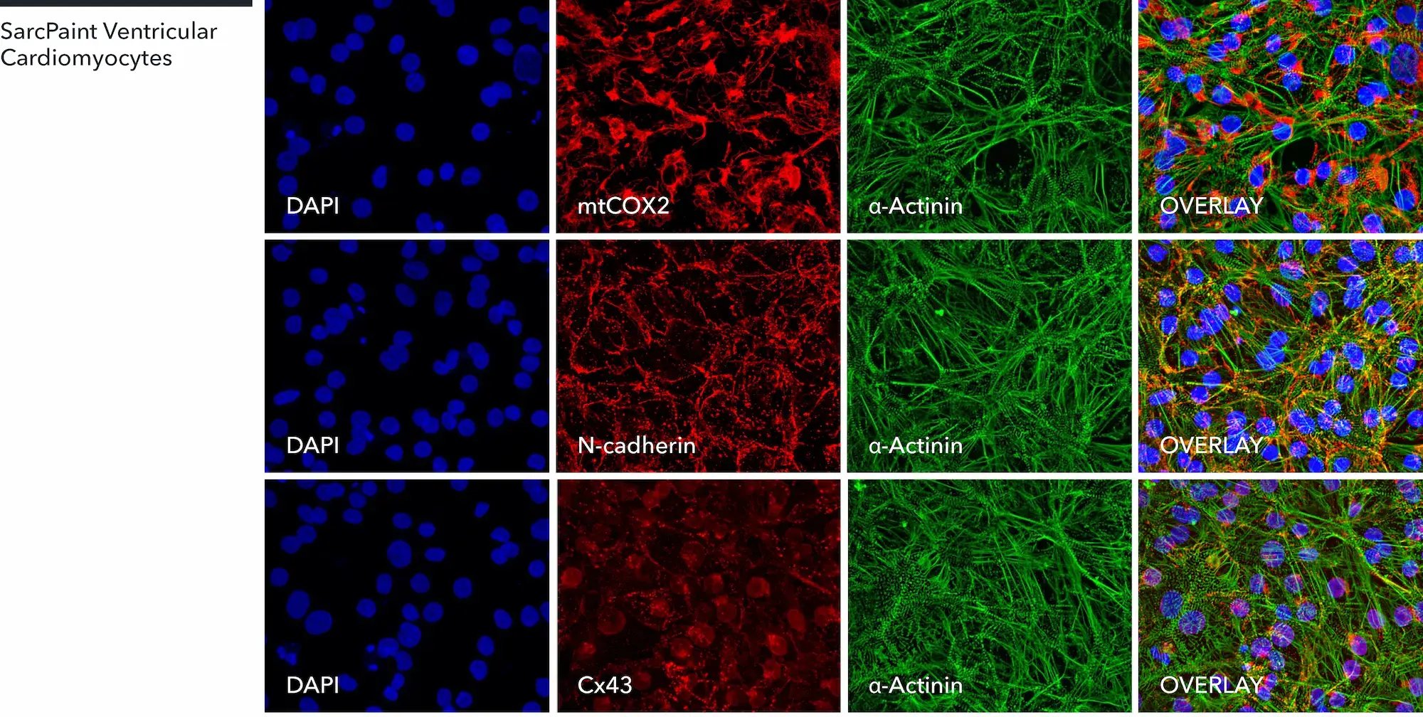
SarcPaint Ventricular Cardiomyocytes express key cardiac specific markers

Immunofluorescent staining of human iPSC-derived SarcPaint Ventricular Cardiomyocytes at 10 days post-thawing showing SarcPaint-tagged α-actinin (green) with co-localisation of mitochondria (mtCOX2), N-cadherin or connexin 43 (Cx43); 60x magnification.

myrcell-ventricular-cardiomyocyte-sarcpaint-sarcomere-remodelling

Live tracking of drug-induced sarcomere remodelling

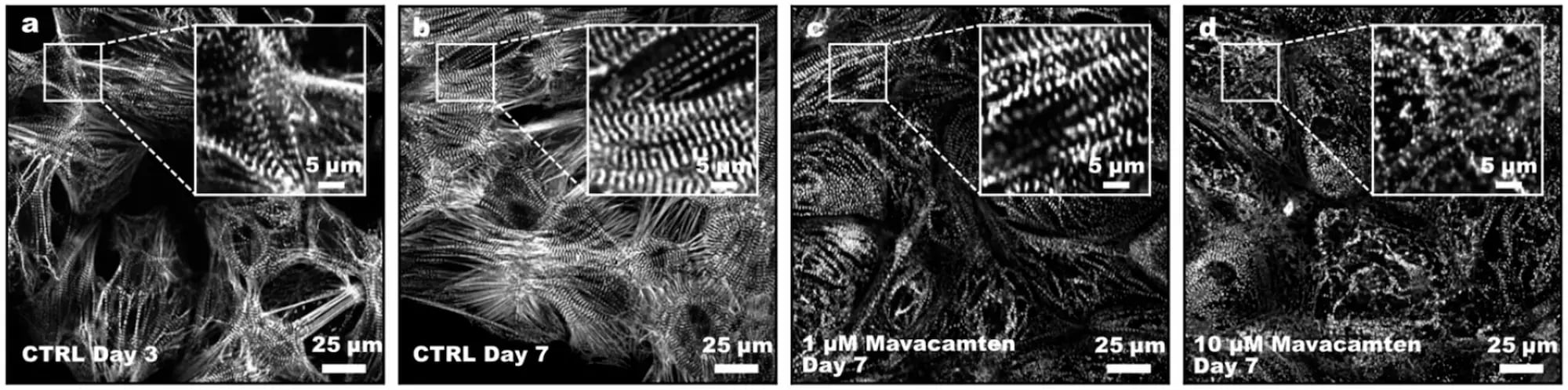
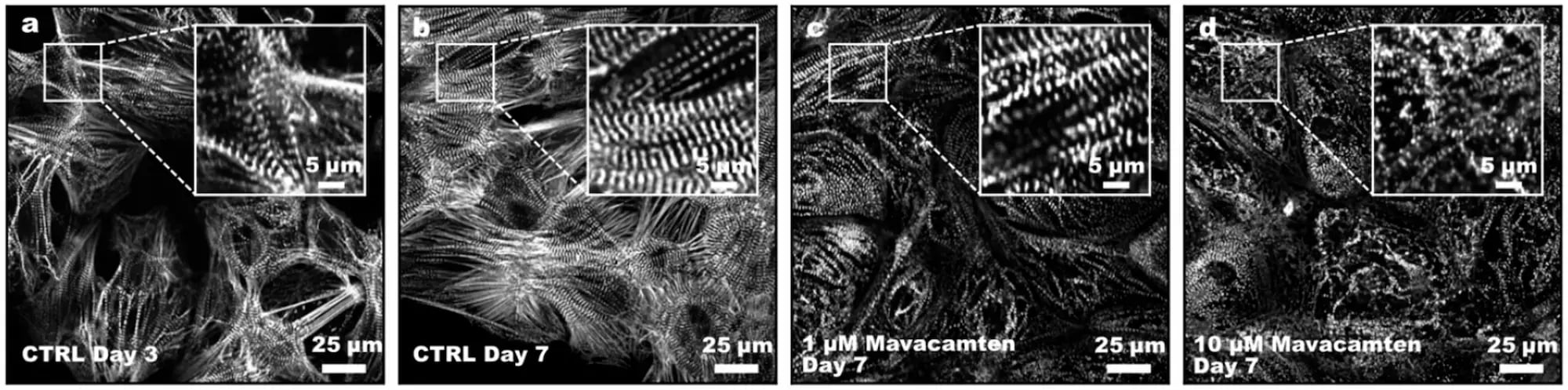
Representative images show live-cell imaging of sarcomere remodelling following mavacamten exposure. SarcPaint Ventricular Cardiomyocytes were imaged during culture and following treatment with mavacamten.

Control cardiomyocytes at days 3 and 7 show progressive organisation of sarcomeric Z-bands (images a and b); treatment with 1 and 10 µM mavacamten at day 7 produces concentration-dependent alterations in sarcomere architecture, with increasing disruption of the organised Z-band pattern at higher exposure (c and d).

Live tracking of drug-induced sarcomere remodelling

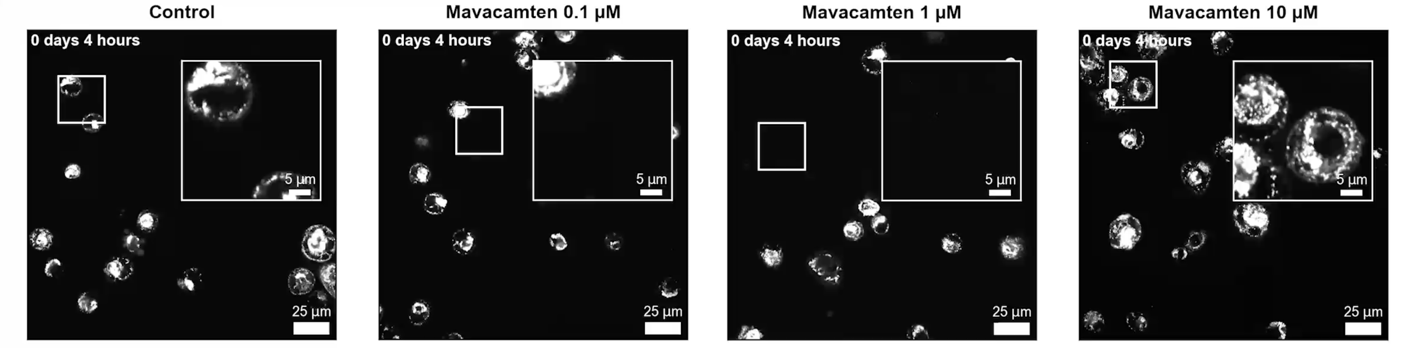
Representative video show live-cell imaging of sarcomere remodelling following mavacamten exposure. SarcPaint Ventricular Cardiomyocytes were imaged during culture and following treatment with mavacamten.

The video shows real-time time-lapse of sarcomeric changes on compound exposure. The endogenous ACTN2-Citrine reporter (SarcPaint) enables direct visualisation of these structural changes in live cardiomyocytes, providing a non-invasive approach for longitudinal assessment of drug-induced sarcomere remodelling and contractile phenotypes.

Control cardiomyocytes at days 3 and 7 show progressive organisation of sarcomeric Z-bands (images a and b); treatment with 1 and 10 µM mavacamten at day 7 produces concentration-dependent alterations in sarcomere architecture, with increasing disruption of the organised Z-band pattern at higher exposure (c and d).

myrcell-ventricular-cardiomyocytes-sarcpaint-compound-screen

Targeted small-scale screen for multiparametric functional insights

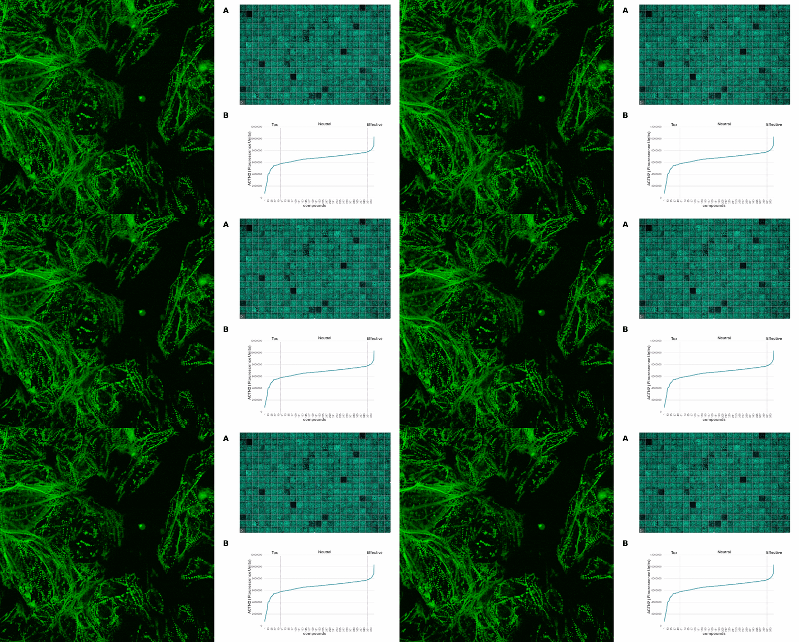
Representative small-scale phenotypic screening workflow using SarcPaint Ventricular Cardiomyocytes. Cells were seeded into 384-well imaging plates and cultured for 10 days post-thaw before compound treatment (A).

Live cardiomyocytes were screened against a targeted library of 377 calcium channel-modulating compounds and imaged using the CellVoyager CV8000 High-Content Screening System (Yokogawa) (left).

AI-assisted image analysis quantified multiple structural and functional phenotypic parameters, including sarcomere organisation, myofibrillar integrity, cell morphology, and contractile behaviour. Compounds were subsequently classified based on their effects on α-actinin-positive sarcomeric architecture, enabling discrimination of beneficial, neutral, and toxic phenotypes and prioritisation of candidate molecules for downstream functional validation (B).

myrcell-ventricular-cardiomyocytes-sarcpaint-phenotypic-screening

Live quantification of contractile responses to reference compounds

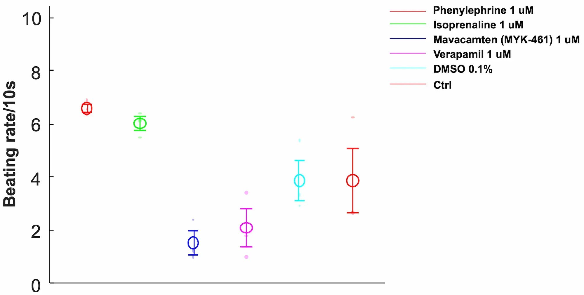
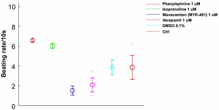
Representative pharmacological characterisation of SarcPaint Ventricular Cardiomyocytes. Cells were cultured on Matrigel-coated plates for 10 days post-thaw and treated with reference compounds targeting distinct cardiac signalling pathways. Live imaging was used to quantify beating rate.

Positive chronotropic agents, including phenylephrine (red) and isoprenaline (green) increased beating frequency, whereas mavacamten (blue) and verapamil (pink) reduced contractile amplitude and beating activity.

These data demonstrate that SarcPaint Ventricular Cardiomyocytes are sensitive to pharmacological modulation, confirming their suitability for functional drug screening.

Tracking functional and structural maturation in 3D engineered heart muscle

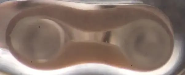
SarcPaint Ventricular Cardiomyocytes were combined with fibroblasts in a collagen-based hydrogel and cast into myrPlates to generate Engineered Heart Muscle (EHM). Cardiomyocytes were maintained for 7-10 days post thaw culture before tissue casting. Live video-optical tracking using automated pole detection and peak analysis in the myrImager platform enabled longitudinal assessment of the tissue contractility throughout maturation.

Representative video of EHM contractility at day 35 of culture.

myrcell-ventricular-cardiomyocytes-sarcpaint-ehm-protein-expression

Tracking functional and structural maturation in 3D engineered heart muscle

SarcPaint Ventricular Cardiomyocytes were combined with fibroblasts in a collagen-based hydrogel and cast into myrPlates to generate Engineered Heart Muscle (EHM). Cardiomyocytes were maintained for 7-10 days post-thaw culture before tissue casting. Live video-optical tracking using automated pole detection and peak analysis in the myrImager platform enabled longitudinal assessment of the tissue contractility throughout maturation.

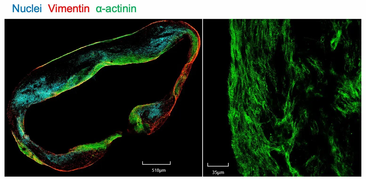
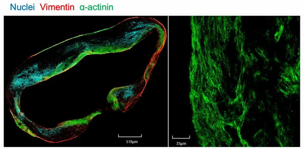
Immunofluorescent imaging of whole EHM mount staining at day 21 (right); a cross section of EHM at day 28 (left). Together the images show well-aligned cardiomyocytes with organised sarcomeres and interspersed fibroblasts.

myrcell-ventricular-cardiomyocytes-sarcpaint-ehm-video-optics-organ-bath

Assessment of contractility

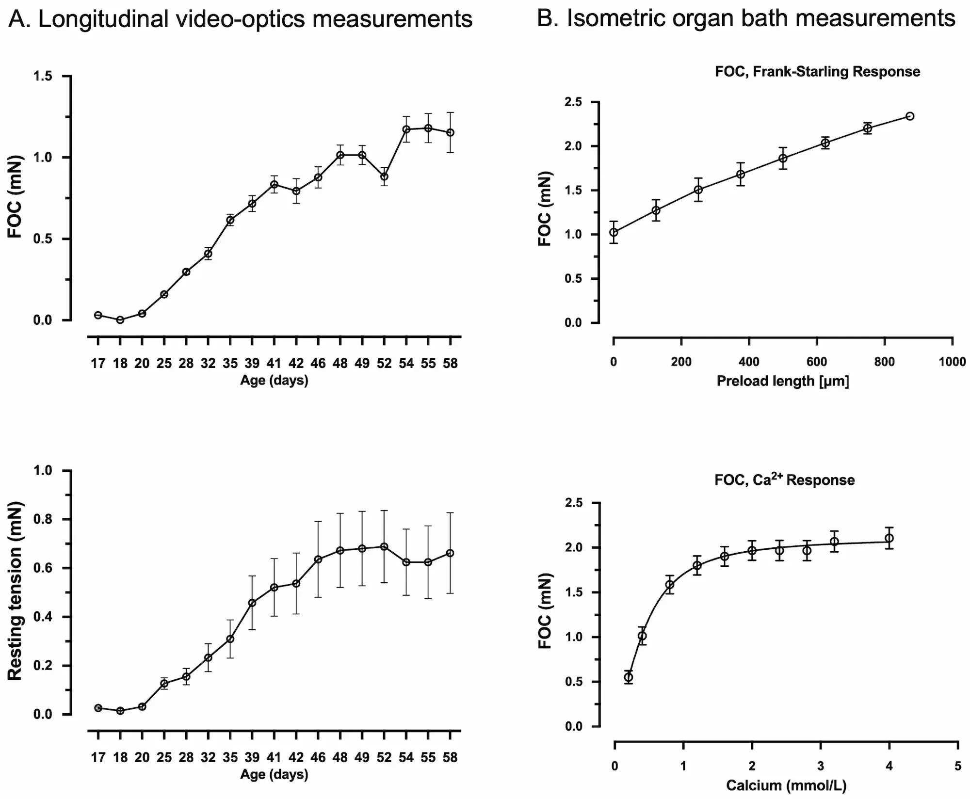
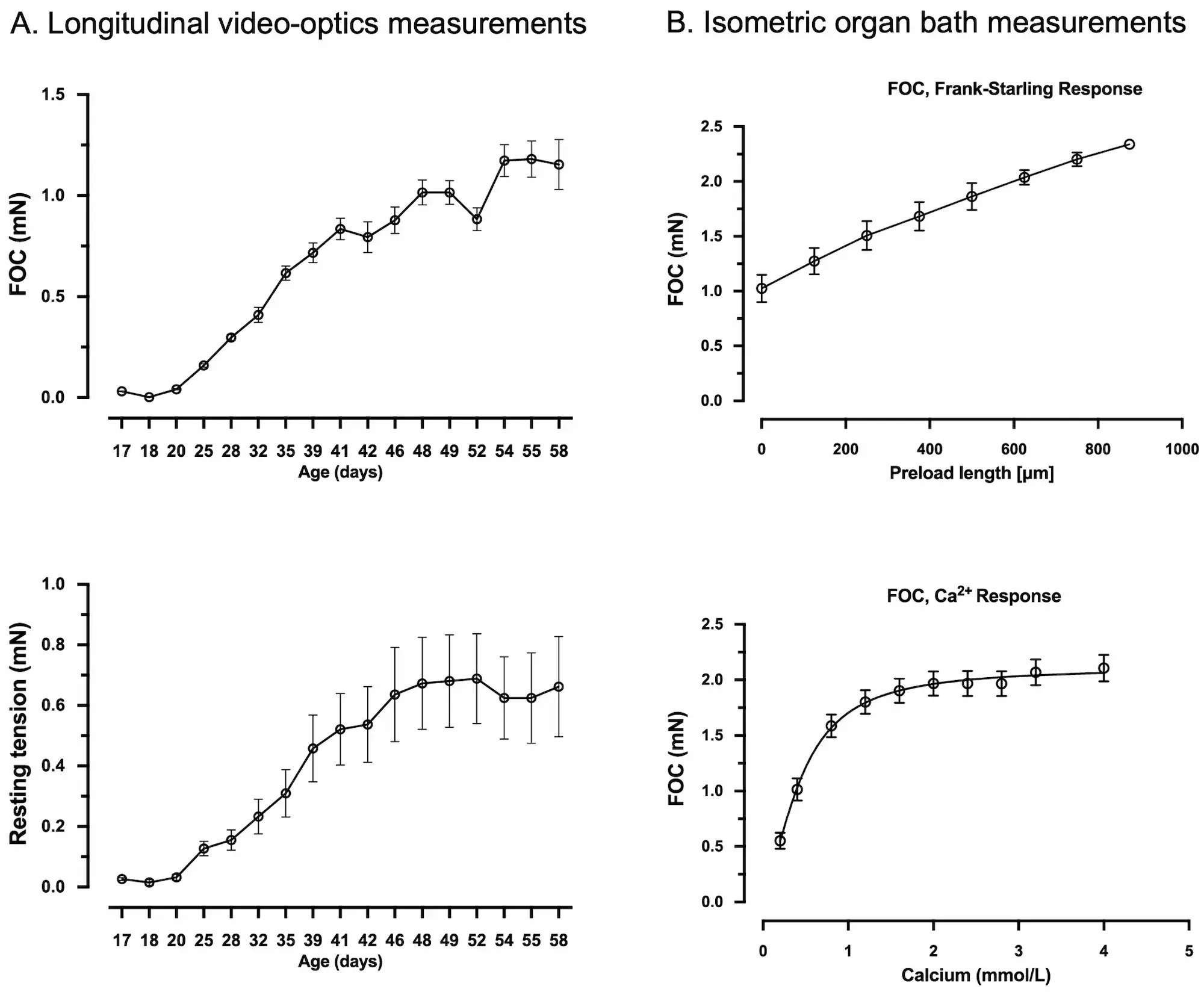
Longitudinal video-optical contractility measurements of 16 tissues over 58 days of culture, demonstrate progressive improvement in force of contraction (FOC) indicative of tissue maturation (A).

Contractile analysis under isometric conditions allowing deep phenotyping of contractility at the study endpoint from tissues at day 58 showing tissue elasticity and increased force development (B).

Data shown as mean ± SEM, n=8.

myrcell-ventricular-cardiomyocyte-ehm-contractility-wt-sarcpaint

Preserved functional response in cells expressing Sarcpaint reporter

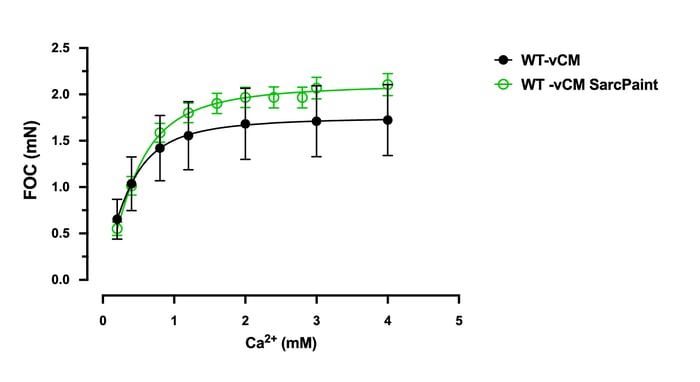
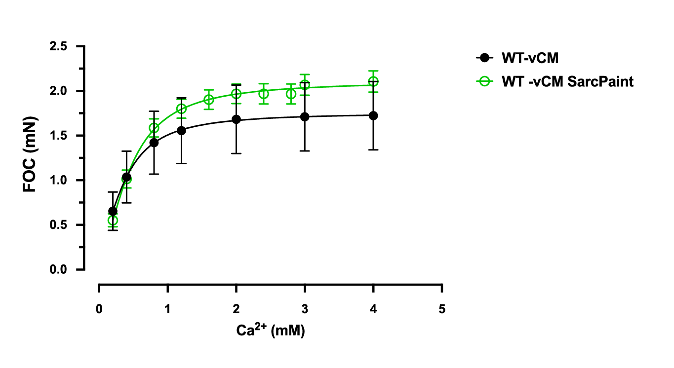
Engineered Heart Muscle (EHM) generated from wild-type Ventricular Cardiomyocytes (WT-vCM) and SarcPaint Ventricular Cardiomyocytes (WT-vCM SarcPaint) show comparable function at similar maturation age. The data shows contractile analysis of WT-vCM at day 42 and WT-vCM SarcPaint at day 58 under isometric conditions. 

Vial limit exceeded

A maximum number of 20 vials applies. If you would like to order more than 20 vials, please contact us at orders@bit.bio.


Human iPSC-derived ventricular cardiomyocytes with SarcPaint reporter

myrCell SarcPaint Ventricular Cardiomyocytes are human iPSC-derived ventricular cardiomyocytes generated using traditional differentiation protocols. The cells carry a genetically encoded ACTN2-Citrine sarcomeric reporter (SarcPaint), delivering live, fluorescently tagged sarcomeric α-actinin for real-time structural readout without fixation or immunostaining. Upon revival, they exhibit a robust ventricular phenotype characterised by the expression of key cardiac markers including cTnT, α-actinin, NCX1, MYL2, MYH7 and MLC2v.

SarcPaint Ventricular Cardiomyocytes form electrically coupled monolayers and exhibit spontaneous, synchronised contractions within 7 days, consistent with the functional timeline of the parental wild-type cells.

Pharmacological characterisation confirms the product's relevance for live functional and compound screening: mavacamten and verapamil reduce contractile force and beating rate, while phenylephrine and isoprenaline increase beating rate. Sarcomere structure and organisation can be quantified using standard image analysis platforms or AI assisted algorithms enabling automated, high-content assessment of sarcomeric organisation analysis directly in live cardiomyocytes.

Ventricular Cardiomyocytes incorporating the SarcPaint reporter provide a tool for cardiotoxicity screening, mechanistic studies of sarcomere biology, and drug discovery programs requiring real-time, quantitative structural readouts alongside contractile function.

Benchtop benefits

quick_0

Live-cell imaging ready

Genetically encoded ACTN2-Citrine reporter enables label-free, real-time visualisation of sarcomere structure without fixation.

functional_0

Functional

Spontaneously beating with ventricular electrophysiology, responsive to cardiac-active compounds in live functional assays.

efficient2_0

Automation compatible

Compatible with image analysis platforms for automated, high-content quantification of sarcomere organisation.

Cells arrive ready to plate

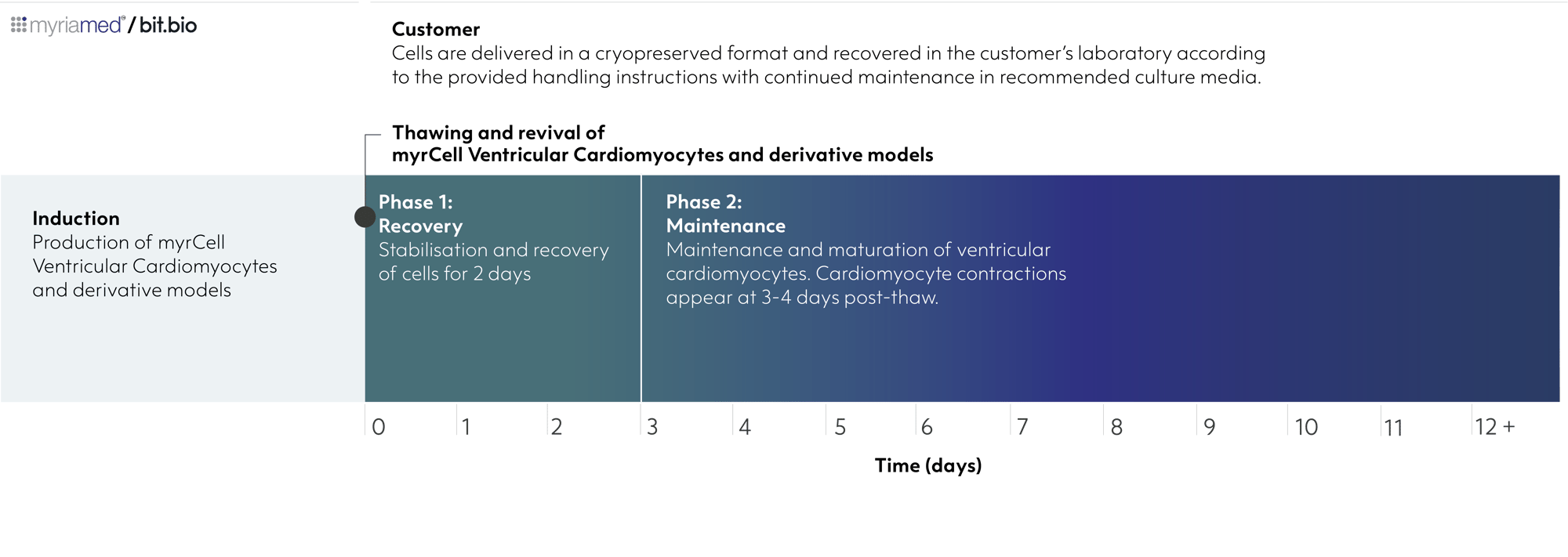
myrcell-ventricular-cardiomyocytes-derivatives-timelineSarcPaint Ventricular Cardiomyocytes are delivered in a cryopreserved format and recovered in the customer’s laboratory according to the provided handling instructions with continued maintenance in recommended culture media.

Product specifications

Starting material

Human iPSC line, W001

Karyotype

Normal (46, XY)

Seeding compatibility

6, 12, 24, 48, 96 & 384 well plates

Shipping info

Dry ice

Donor

Male

Vial size

Small: >1 x 10 viable cells

Quality control

Sterility, protein expression (ICC)

Differentiation method

Small molecule-directed differentiation

Recommended minimum seeding density

90,000 cells/cm² (laminin-coated plates); 150,000 cells/cm² (Matrigel-coated plates)

User storage

LN2 or -150°C

Format

Cryopreserved cells

Genetic modification

SarcPaint: genetically encoded ACTN2-Citrine sarcomeric reporter

Applications

Cardiovascular research
Disease modelling
2D screening and functional assays
3D engineered heart muscle

Product use

myrCells are for research use only


Scale your study with volume pricing

Enabling scientists to use human cells in their research, running additional experiments without rationing cells or limiting experimental scale


Order quantity Total vials received Pricing tier 
1 - 9 packs 3 - 27 vials Standard price
10 - 33 packs 30 - 99 vials Automatic 10% discount
> 34 packs > 100 vials > Contact us for a quote


Technical data

SarcPaint reporter enables real-time functional assessment

Real-time imaging of sarcomeric dynamics in SarcPaint Ventricular Cardiomyocytes

Live fluorescent recording of SarcPaint Ventricular Cardiomyocytes, cultured in Matrigel-coated flasks; recorded at Day 10 post-thaw. Scale: 100 μm, 60x magnification.

Real-time monitoring and longitudinal analysis of sarcomere organisation and function

Automated extraction of Z-band profiles and quantitative measurement of sarcomere spacing from confocal images enabling deep learning-based segmentation and spatial mapping with automated AI assisted workflows. This model enables automated multi-scale quantification of sarcomere organisation, myofibril architecture, and contractile dynamics directly from live-cell images and videos, thus eliminating endpoint staining while supporting longitudinal phenotyping and high-content drug screening.

Highly characterised

SarcPaint Ventricular Cardiomyocytes express key cardiac specific markers

myrcell-sarcpaint-ventricular-cardiomyocytes-icc-ctnt-ctni-mlc2v

Immunofluorescent staining of human iPSC-derived SarcPaint Ventricular Cardiomyocytes at 10 days post-thawing showing SarcPaint-tagged α-actinin (green) with co-localisation of cardiac troponin T (cTnT), cardiac troponin I (cTnI) or ventricular myosin light chain-2 (MLC2v); 60x magnification.

myrcell-sarcpaint-ventricular-cardiomyocytes-icc-mtcox2-n-cadherin-cx43
Immunofluorescent staining of human iPSC-derived SarcPaint Ventricular Cardiomyocytes at 10 days post-thawing showing SarcPaint-tagged α-actinin (green) with co-localisation of mitochondria (mtCOX2), N-cadherin or connexin 43 (Cx43); 60x magnification.
Technical data

SarcPaint reporter enables real-time functional assessment

Real-time imaging of sarcomere dynamics in SarcPaint Ventricular Cardiomyocytes

Live fluorescent recording of SarcPaint Ventricular Cardiomyocytes, cultured in Matrigel-coated flasks; recorded at Day 10 post-thaw. Scale: 100 μm, 60x magnification.

Real-time monitoring and longitudinal analysis of sarcomere organisation and function

Automated extraction of Z-band profiles and quantitative measurement of sarcomere spacing from confocal images enabling deep learning-based segmentation and spatial mapping with automated AI assisted workflows. This model enables automated multi-scale quantification of sarcomere organisation, myofibril architecture, and contractile dynamics directly from live-cell images and videos, thus eliminating endpoint staining while supporting longitudinal phenotyping and high-content drug screening.

Live tracking of drug-induced sarcomere remodelling

myrcell-ventricular-cardiomyocyte-sarcpaint-sarcomere-remodellingRepresentative video (upper panel) and images (lower panel) show live-cell imaging of sarcomere remodelling following mavacamten exposure. SarcPaint Ventricular Cardiomyocytes were imaged during culture and following treatment with mavacamten.
The video shows real-time time-lapse of sarcomeric changes on compound exposure. The endogenous ACTN2-Citrine reporter (SarcPaint) enables direct visualisation of these structural changes in live cardiomyocytes, providing a non-invasive approach for longitudinal assessment of drug-induced sarcomere remodelling and contractile phenotypes.

Control cardiomyocytes at days 3 and 7 show progressive organisation of sarcomeric Z-bands (images a and b); treatment with 1 and 10 µM mavacamten at day 7 produces concentration-dependent alterations in sarcomere architecture, with increasing disruption of the organised Z-band pattern at higher exposure (c and d).

Phenotypic screening

Targeted small-scale screen for multiparametric functional insights

myrcell-ventricular-cardiomyocytes-sarcpaint-compound-screen

Representative small-scale phenotypic screening workflow using SarcPaint Ventricular Cardiomyocytes. Cells were seeded into 384-well imaging plates and cultured for 10 days post-thaw before compound treatment (A).

Live cardiomyocytes were screened against a targeted library of 377 calcium channel-modulating compounds and imaged using the  CellVoyager CV8000 High-Content Screening System (Yokogawa) (left).

AI-assisted image analysis quantified multiple structural and functional phenotypic parameters, including sarcomere organisation, myofibrillar integrity, cell morphology, and contractile behaviour. Compounds were subsequently classified based on their effects on α-actinin-positive sarcomeric architecture, enabling discrimination of beneficial, neutral, and toxic phenotypes and prioritisation of candidate molecules for downstream functional validation (B).

Live quantification of contractile responses to reference compounds

myrcell-ventricular-cardiomyocytes-sarcpaint-phenotypic-screening

Representative pharmacological characterisation of SarcPaint Ventricular Cardiomyocytes. Cells were cultured on Matrigel-coated plates for 10 days post-thaw and treated with reference compounds targeting distinct cardiac signalling pathways. Live imaging was used to quantify beating rate.

Positive chronotropic agents, including phenylephrine (red) and isoprenaline (green) increased beating frequency, whereas mavacamten (blue) and verapamil (pink) reduced contractile amplitude and beating activity.

These data demonstrate that SarcPaint Ventricular Cardiomyocytes are sensitive to pharmacological modulation, confirming their suitability for functional drug screening.

Functional engineered cardiac tissues

Tracking functional and structural maturation in 3D engineered heart muscle

myrcell-ventricular-cardiomyocytes-sarcpaint-ehm-protein-expression

SarcPaint Ventricular Cardiomyocytes were combined with fibroblasts in a collagen-based hydrogel and cast into myrPlates to generate Engineered Heart Muscle (EHM). Cardiomyocytes were maintained for 7-10 days post thaw culture before tissue casting. Live video-optical tracking using automated pole detection and peak analysis in the myrImager platform enabled longitudinal assessment of the tissue contractility throughout maturation.

Representative video of EHM contractility at day 35 of culture (upper panel).

Immunofluorescent imaging of whole EHM mount staining at day 21 (lower panel, right); a cross section of EHM at day 28 (left). Together the images show well aligned cardiomyocytes with organised sarcomeres and interspersed fibroblasts.

Assessment of contractility

myrcell-ventricular-cardiomyocytes-sarcpaint-ehm-video-optics-organ-bath

Longitudinal video-optical contractility measurements of 16 tissues over 58 days of culture, demonstrate progressive improvement in force of contraction (FOC) indicative of tissue maturation (A).

Contractile analysis under isometric conditions allowing deep phenotyping of contractility at the study endpoint from tissues at day 58 showing tissue elasticity and increased force development (B).

Data shown as mean ± SEM, n=8.

Preserved functional response in cells expressing Sarcpaint reporter

myrcell-ventricular-cardiomyocyte-ehm-contractility-wt-sarcpaint

Engineered Heart Muscle (EHM) generated from wild-type Ventricular Cardiomyocytes (WT-vCM) and SarcPaint Ventricular Cardiomyocytes (WT-vCM SarcPaint) show comparable function at similar maturation age. The data shows contractile analysis of WT-vCM at day 42 and WT-vCM SarcPaint at day 58 under isometric conditions.

Frequently Asked Questions (FAQs)

What is the SarcPaint reporter?

SarcPaint is a genetically encoded ACTN2-Citrine knock-in reporter that fluorescently tags sarcomeric α-actinin, enabling live, label-free imaging and quantification of sarcomere structure and organisation in cardiomyocytes without fixation or immunostaining (Haertter D. et al., 2025).


Does the SarcPaint tag affect cardiomyocyte function?

The parental ventricular cardiomyocyte line was engineered with the genetically encoded ACTN2-Citrine knock-in reporter (SarcPaint). The resulting derivative, SarcPaint Ventricular Cardiomyocytes, fully retains the defined ventricular phenotype and robust functional performance of the unmodified parental cells.


Can SarcPaint-engineered cardiomyocytes be used for compound screening?

SarcPaint Ventricular Cardiomyocytes are well-suited for compound screening; pharmacological characterisation shows that mavacamten and verapamil reduce contractile force and beating rate, while phenylephrine and isoprenaline increase beating rate, confirming their pharmacological relevance for contractility and calcium-channel-modulator assays.


Is fixation required to visualise sarcomere structure?

The ACTN2-Citrine (SarcPaint) reporter allows live, label-free imaging of sarcomere assembly and organisation over time, without the need for fixation or immunostaining.


How long can SarcPaint Ventricular Cardiomyocytes be maintained in culture?

SarcPaint Ventricular Cardiomyocytes can be maintained in culture for up to 8 weeks while retaining stable electrophysiological properties, ventricular identity, and contractile function. Similar to the wild-type parental product, they establish electrically coupled monolayers and begin spontaneous, synchronised contractions within 3–7 days post-thaw.


What applications are SarcPaint Cardiomyocytes suitable for?

SarcPaint Ventricular Cardiomyocytes are compatible with live-cell sarcomere imaging, contractility and cardiotoxicity assessments, compound screening, and structural analysis via the SarcAsM platform (Haertter D. et al., 2025), in addition to all standard applications of the unmodified parental Ventricular Cardiomyocytes.


ioCells catalogue

Human iPSC-derived cells

powered by opti-ox

Consistent. Defined. Scalable.

bitbio-cell_catalogue_header-with-tracker-Desktop-2500x1664