myrCell Ventricular Cardiomyocytes
myrcell-ventricular-cardiomyocytes-alpha-actinin-tnni3
myrCell-ventricular-cardiomyocytes-synchronised-beating
myrcell-ventricular-cardiomyocytes-morphology
myrcell-ventricular-cardiomyoctyes-protein-expression-ctnt-mtcox2
myrcell-cardiomyocytes-rna-seq-ventricular-atrial-phenotypes
myrcell-ventricular-atrial-cardiomyocytes-patch-clamp-ion-channel-activity
myrcell-ventricular-atrial-cardiomyocytes-patch-clamp-action-potential
myrcell-ventricular-atrial-cardiomyocytes-contraction-cytomotion
myrcell-ventricular-cardiomyocytes-mitochondrial-function
myrcell-cardiomyocytes-ehm-contraction-thumbnail
myrcell-cardiomyocytes-ehm-protein-expression-mito-actinin
myrcell-ventricular-cardiomyocytes-ehm-force-of-contraction
myrcell-ventricular-cardiomyocytes-ehm-metabolic-flexibility-oxidative-maturation
myrcell-ventricular-cardiomyocytes-alpha-actinin-tnni3
myrCell-ventricular-cardiomyocytes-synchronised-beating
myrcell-ventricular-cardiomyocytes-morphology
myrcell-ventricular-cardiomyoctyes-protein-expression-ctnt-mtcox2
myrcell-cardiomyocytes-rna-seq-ventricular-atrial-phenotypes
myrcell-ventricular-atrial-cardiomyocytes-patch-clamp-ion-channel-activity
myrcell-ventricular-atrial-cardiomyocytes-patch-clamp-action-potential
myrcell-ventricular-atrial-cardiomyocytes-contraction-cytomotion
myrcell-ventricular-cardiomyocytes-mitochondrial-function
myrcell-cardiomyocytes-ehm-contraction-thumbnail
myrcell-cardiomyocytes-ehm-protein-expression-mito-actinin
myrcell-ventricular-cardiomyocytes-ehm-force-of-contraction
myrcell-ventricular-cardiomyocytes-ehm-metabolic-flexibility-oxidative-maturation

cat no | 02-1001-0000

myrCell Ventricular Cardiomyocytes

Human iPSC-derived ventricular cardiomyocytes

  • Cryopreserved human iPSC-derived ventricular cardiomyocytes generated by directed differentiation that are ready for experiments in days

  • Ideal for studying cardiovascular disease and cardiotoxicity in 2D or 3D engineered heart muscle

  • Highly characterised to show functional synchronised beating by day 7 post-revival
myrcell-ventricular-cardiomyocytes-alpha-actinin-tnni3

Human iPSC-derived ventricular cardiomyocytes

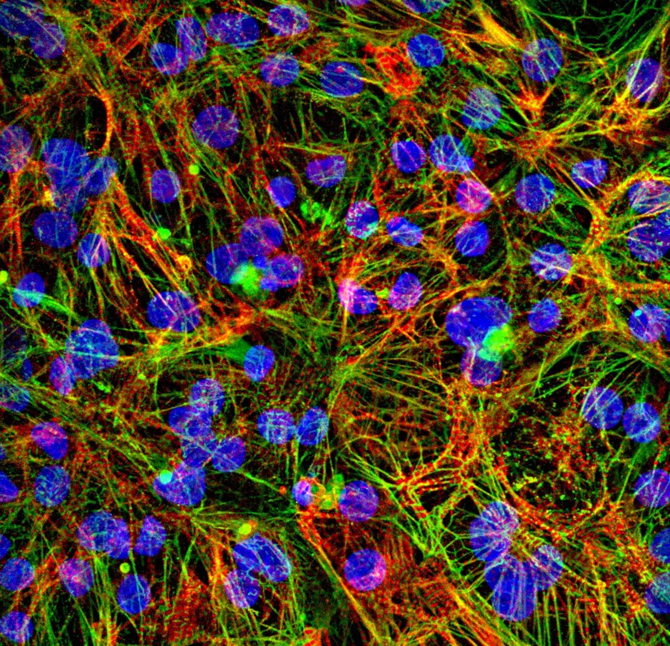
Immunofluorescent staining of human iPSC-derived Ventricular Cardiomyocytes 10 days post-thawing showing expression of α-actinin (structural protein in the Z-discs, responsible for organised contractile sarcomere; green), and cardiac troponin I (TNNI3) (a cardiac-specific component of the troponin complex involved in regulation of myocardial contraction and a marker of differentiated ventricular cardiomyocytes; red) and nuclei stained with DAPI (blue). 60x magnification.

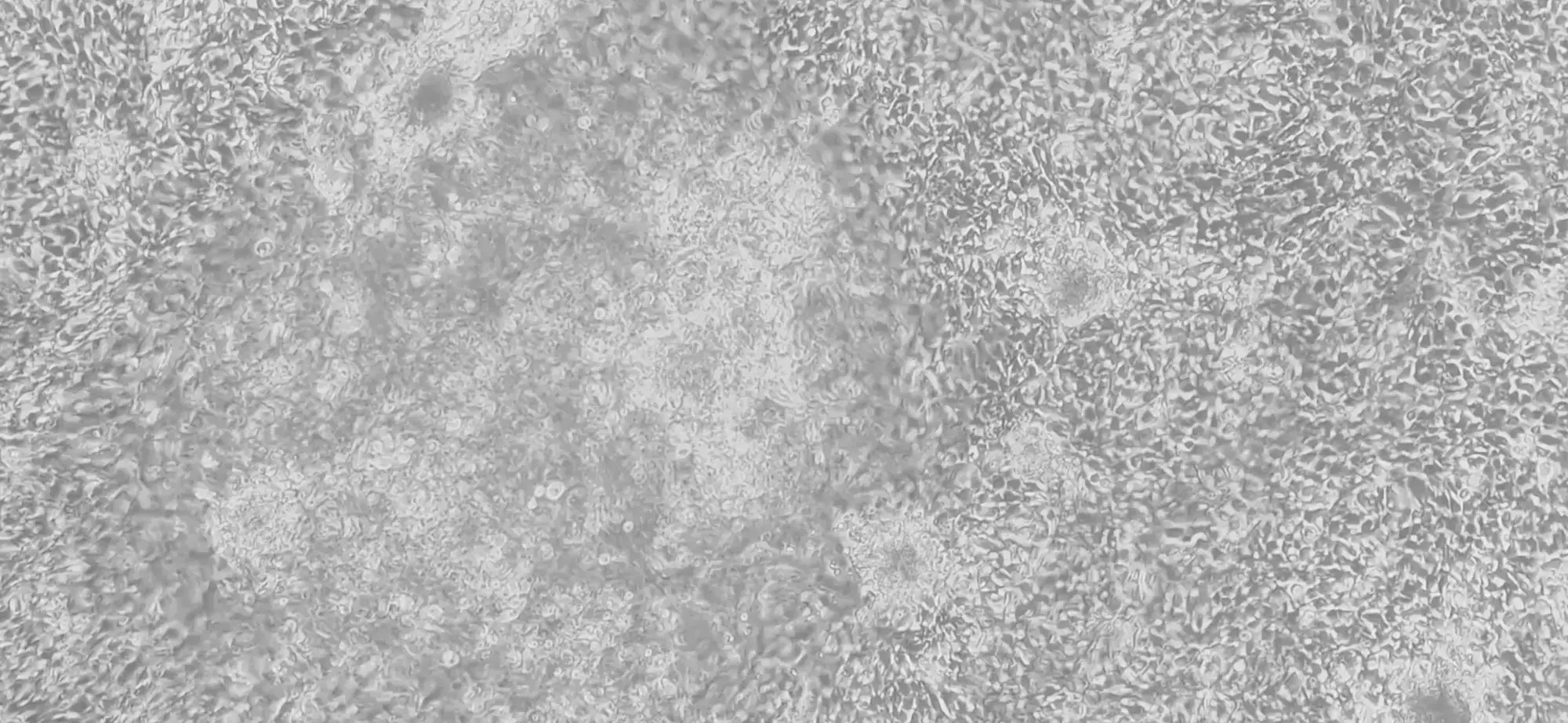
Ventricular Cardiomyocytes show spontaneous, synchronised beating by day 7

Representative brightfield video showing spontaneous, synchronised beating of Ventricular Cardiomyocytes cultured in Matrigel-coated flasks at day 7 post-thaw; 4x magnification. 

myrcell-ventricular-cardiomyocytes-morphology

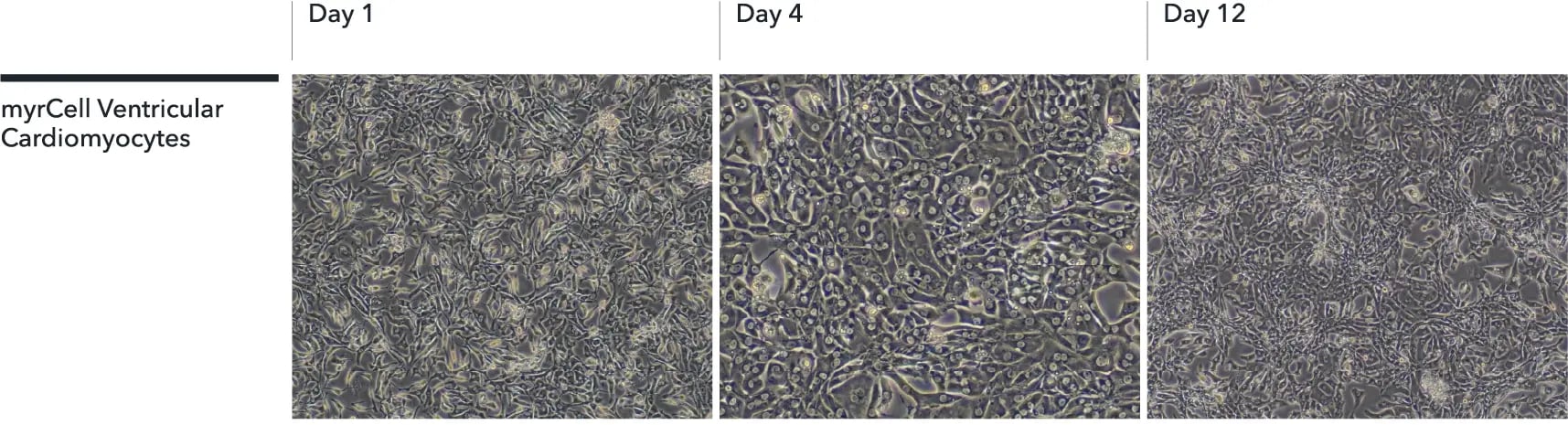
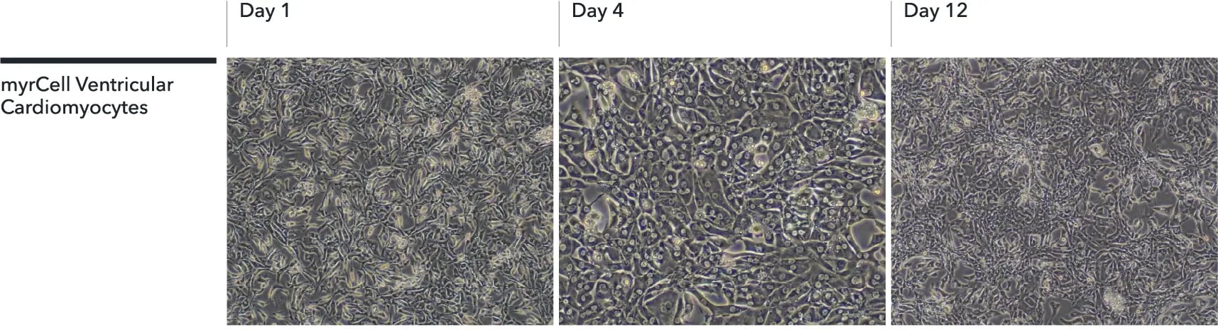
Ventricular Cardiomyocytes form confluent, electrically coupled monolayers by day 12

Brightfield images showing the progressive recovery and attachment of Ventricular Cardiomyocytes cultured on Matrigel-coated flasks. Cells form confluent, electrically coupled monolayers and develop spontaneous, synchronised beating following thawing. Images were acquired at days 1, 4, and 12 post-thaw (10x objective). 

myrcell-ventricular-cardiomyoctyes-protein-expression-ctnt-mtcox2

Ventricular Cardiomyocytes express key cardiac specific markers

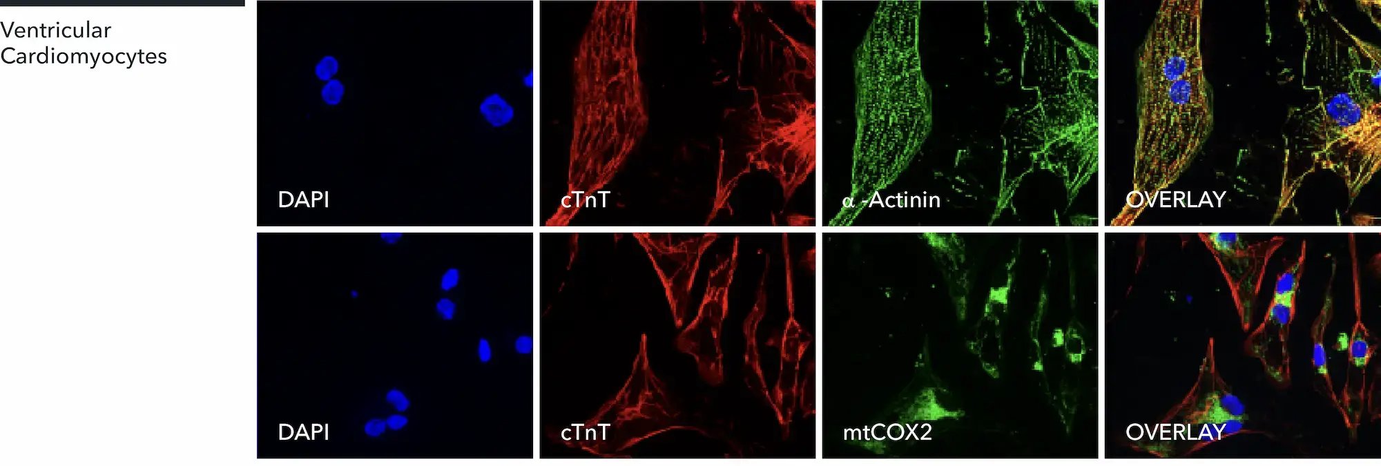
Representative immunofluorescent staining of human iPSC-derived ventricular cardiomyocytes at 10 days post-thaw showing co-localisation of cardiac troponin T (cTnT) with α-actinin, and cTNT with mitochondria (mtCOX2); 60x magnification.

myrcell-cardiomyocytes-rna-seq-ventricular-atrial-phenotypes

RNA sequencing confirms distinct ventricular and atrial phenotypes

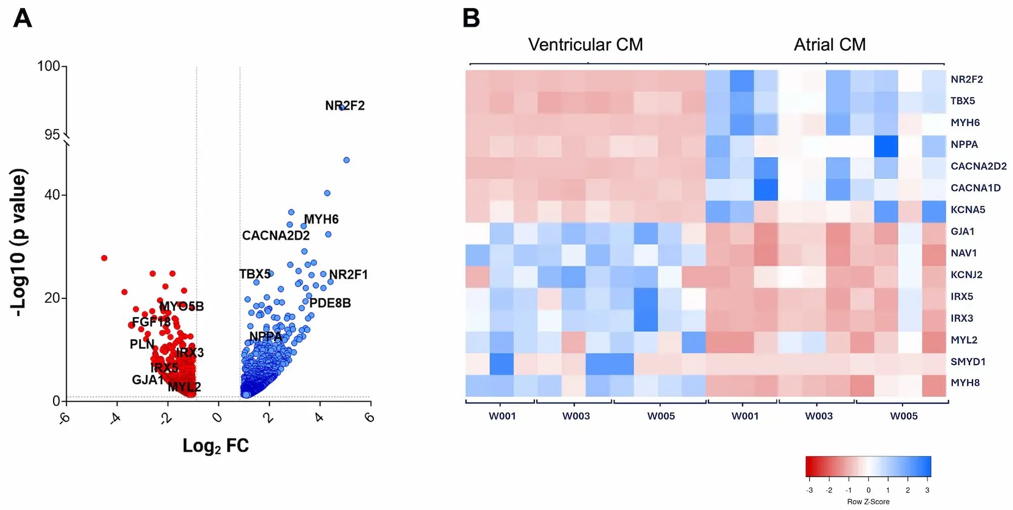
RNA sequencing (RNA-seq) demonstrates robust chamber-specific differentiation of Ventricular and Atrial Cardiomyocytes (CM). Differential gene expression and hierarchical clustering reveal distinct ventricular and atrial transcriptomic signatures, confirming reproducible lineage specification and molecular identity across independent differentiation batches. The cells were harvested for RNA-seq at day 7 post-thaw.

Volcano plot showing differential gene expression. The x-axis represents log2 fold change (FC) and the y-axis shows -log10 (p value) for each gene. Genes with significant upregulation (blue) or downregulation (red) are labeled, with prominent cardiac-related genes (A).

Heatmap depicting the relative expression (row Z-score) of selected cardiac-related genes across samples; gene names are listed on the right. The heatmap reveals distinct clustering patterns between conditions. The colour scale ranges from -3 (red, indicating lower expression) to +3 (blue, indicating higher expression) (B).

Data were generated from three wild-type iPSC lines (W001, W003, W005) differentiated into both ventricular and atrial cardiomyocytes across 3–4 independent batches. For each line and batch, three technical replicates were pooled prior to sequencing. Ventricular Cardiomyocytes, cat. no. 02-1001-0000, and Atrial Cardiomyocytes, cat. no. 06-1001-0000, are generated from iPSC line W001. Please enquire for further information about iPSC lines W003 and W005.

myrcell-ventricular-atrial-cardiomyocytes-patch-clamp-ion-channel-activity

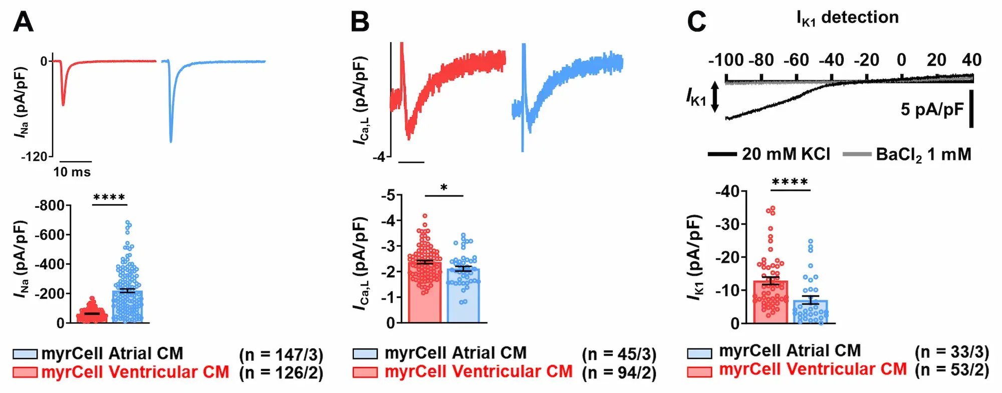
Robust functional expression of key cardiac ion channels confirmed by automated patch-clamp analysis

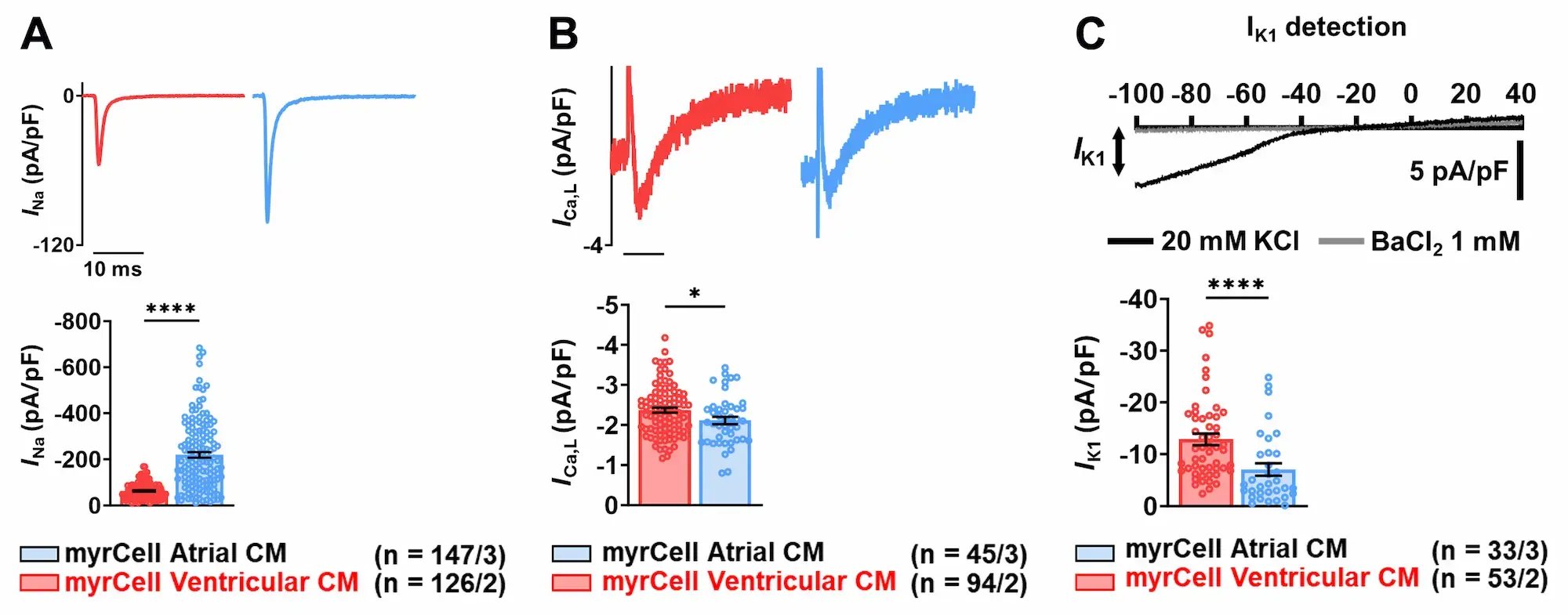
Automated patch-clamp analysis confirms robust expression of key cardiac ion channels in Ventricular (red) and Atrial (blue) Cardiomyocytes (CM).

Representative INa current traces with mean ± SEM peak INa amplitude and I-V curve (A).

Representative ICa,L current traces with mean ± SEM peak ICa,L amplitude and I-V curve fitted with Boltzmann-Ohm curve (B).

Representative trace showing basal inward rectifier current (IK1) in the presence of 20 mM KCl (black) and BaCl2 (grey) and mean ± SEM IK1 at -100 mV (C).

Measurements were acquired from iPSC-derived cardiomyocytes cultured for 50 days post-thaw. Comparisons were made using unpaired Student’s t-test, Mann Whitney’s U-test vs. Ventricular. ****p<0.0001, *p<0.05; n/N = iPSC-CM/batches. 

myrcell-ventricular-atrial-cardiomyocytes-patch-clamp-action-potential

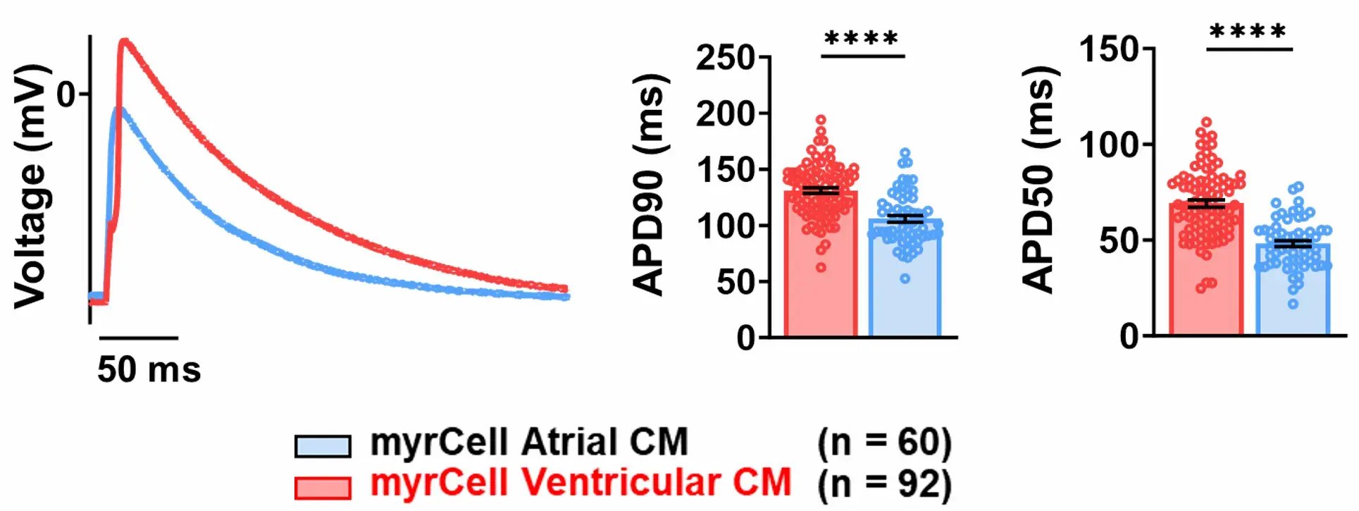
Mature electrophysiological phenotypes demonstrated by action potential recordings

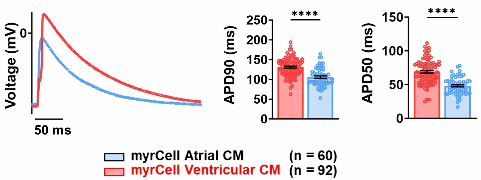
Representative action potential (AP) recordings demonstrate mature ventricular and atrial phenotypes.

Representative membrane voltage (mV) traces showing atrial and ventricular triggered APs in Atrial (blue) and Ventricular (red) Cardiomyocytes (CM) (left).

AP duration at 90% (APD90) and 50% (APD50) repolarisation as mean ±SEM (centre and right).

Measurements were acquired from human iPSC-derived cardiomyocytes cultured for 7 days post-thaw. Comparisons were made using unpaired Student's t-test, Mann Whitney's U-test vs. Ventricular. ****p<0.0001; n = iPSC-CM from one batch. 

myrcell-ventricular-atrial-cardiomyocytes-contraction-cytomotion

Chamber-specific contractile properties confirmed by quantitation of contractile dynamics

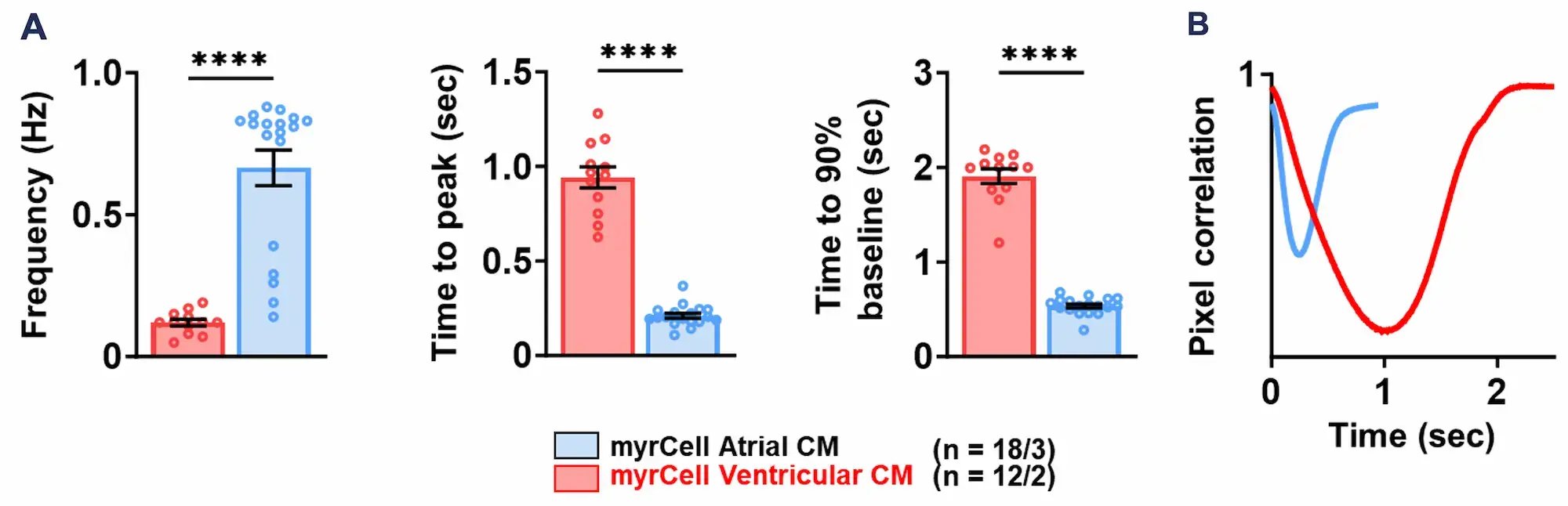
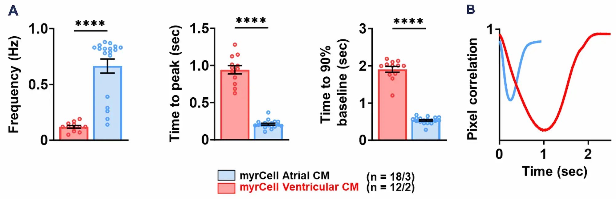
Contraction analysis using CytoMotion label-free, image-based motion detection software (IonOptix), confirms chamber-specific contractile function of Ventricular (red) and Atrial (blue) Cardiomyocytes (CM).

Frequency (Hz), time to peak (sec), and time to 90% baseline (sec) showed as mean ± SEM (A).

Representative traces showing contraction of human iPSC-derived cardiomyocytes monolayers as mean ± SEM (B).

Measurements were acquired from iPSC-derived cardiomyocytes cultured for 50 days post-thaw. Comparisons were made using unpaired Student's t-test, Mann Whitney's U-test vs. Ventricular. ****p<0.001; n/N = measurement ROI/batches. 

myrcell-ventricular-cardiomyocytes-mitochondrial-function

Mitochondrial stress test confirms metabolically mature bioenergetics

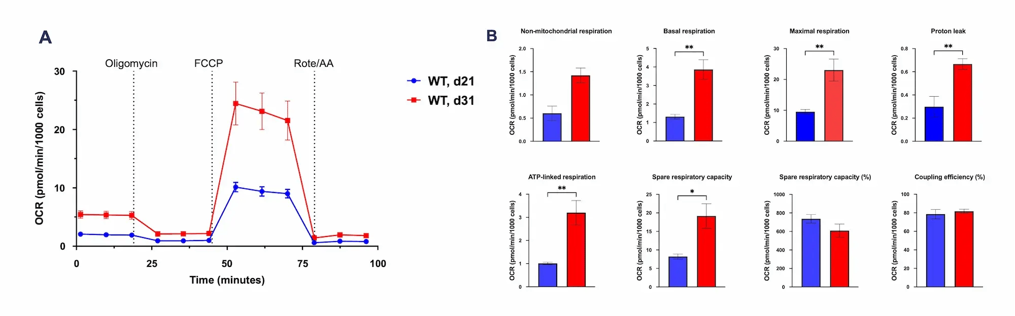
The Seahorse XF Mito Stress Test demonstrates robust mitochondrial respiration and oxidative metabolism in wild-type Ventricular Cardiomyocytes (WT). Cells were maintained in continuous culture and analysed at two maturation stages (Day 21 and Day 31). Seven days prior to the assay (Day 14 and Day 24), cells were seeded into XFe24 cell culture plates at 80,000 cells per well. On the day of the experiment, mitochondrial function was assessed using the Seahorse XF Mito Stress XF RPMI medium, pH 7.4 supplemented with 10 mM Glucose, 1 mM pyruvate, 200 µM L-AsC, 2% B27.

Representative trace of changes in oxygen consumption rate in Ventricular Cardiomyocytes when treated with compounds affecting the mitochondrial electron transport chain, showed as mean ± SEM (A).

Mitochondrial respiration, spare respiratory capacity, ATP production and efficiency derived from the representative trace displayed in (A), shown as mean ± SEM (B).

As cardiomyocytes mature from Day 21 to Day 31, they develop enhanced mitochondrial function and bioenergetic capacity, evidenced by increased mitochondrial respiration, higher ATP-linked respiration, and greater spare respiratory capacity.
Comparisons were made using unpaired Student’s t-test, *p<0.05, **p<0.01, n = 5. 

Engineered heart muscle exhibits progressive maturation and a positive force-frequency relationship

Human iPSC-derived ventricular cardiomyocytes were combined with fibroblasts in a collagen-based hydrogel and cast into myrPlates to generate Engineered Heart Muscle (EHM). Cardiomyocytes were cultured for 7-10 days post thaw before tissue casting. Live video-optical tracking using automated pole detection and peak analysis in the myrImager platform enabled longitudinal assessment of tissue contractility throughout maturation.

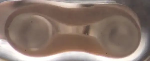
Video-optical recording demonstrates spontaneous tissue contraction and enables non-invasive monitoring of functional maturation.

myrcell-cardiomyocytes-ehm-protein-expression-mito-actinin

Engineered heart muscle exhibits progressive maturation and a positive force-frequency relationship

Human iPSC-derived ventricular cardiomyocytes were combined with fibroblasts in a collagen-based hydrogel and cast into myrPlates to generate Engineered Heart Muscle (EHM). Cardiomyocytes were cultured for 7-10 days post thaw before tissue casting. Live video-optical tracking using automated pole detection and peak analysis in the myrImager platform enabled longitudinal assessment of tissue contractility throughout maturation.

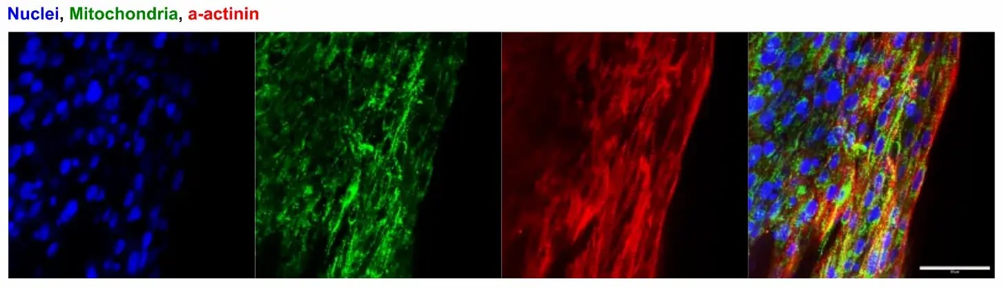
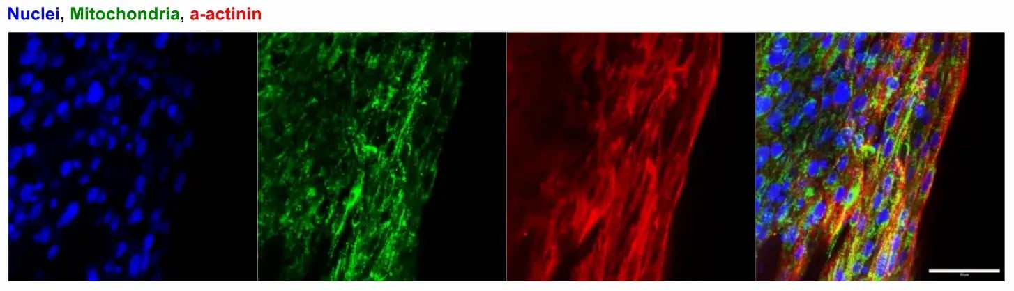
Immunofluorescent imaging of the cross section shows a compact, organised tissue with aligned cardiomyocytes, structured sarcomeres and abundant mitochondria. 

myrcell-ventricular-cardiomyocytes-ehm-force-of-contraction

Engineered heart muscle exhibits progressive maturation and a positive force-frequency relationship

Human iPSC-derived ventricular cardiomyocytes were combined with fibroblasts in a collagen-based hydrogel and cast into myrPlates to generate Engineered Heart Muscle (EHM). Cardiomyocytes were cultured for 7-10 days post thaw before tissue casting. Live video-optical tracking using automated pole detection and peak analysis in the myrImager platform enabled longitudinal assessment of tissue contractility throughout maturation.

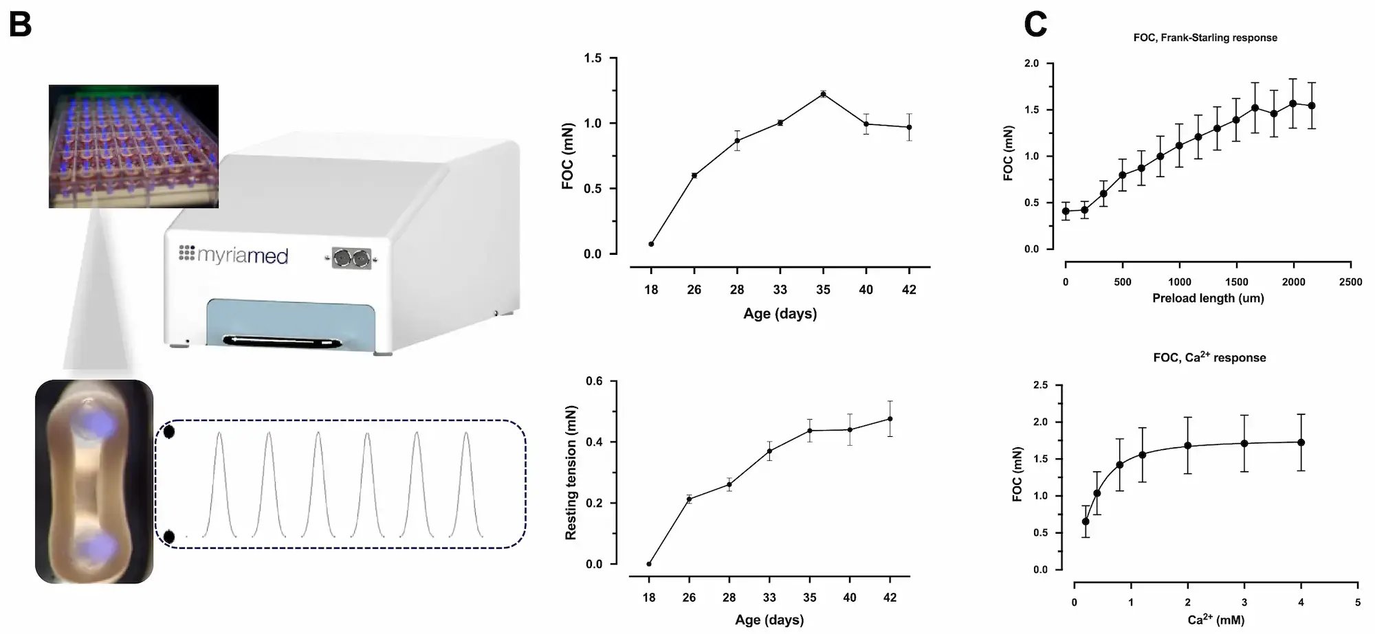
Video-optical measurements of 16 EHMs over 42 days demonstrated progressive increases in force of contraction (FOC), consistent with continued functional maturation (B). Resting length, quantified as the diastolic pole-to-pole distance in myrPlate cultures, provides a longitudinal readout of tissue compaction and mechanical state. Data are shown as mean ± SEM, n=16.

At study endpoint, EHMs were subjected to isometric organ-bath analysis to independently characterise and validate tissue contractile properties (C). The upper panel shows the preload-dependent Frank–Starling response during 1.5 Hz electrical stimulation at 1.8 mM extracellular Ca2+. Stepwise increases in preload assess the relationship between initial muscle length and force development, a fundamental property of functional myocardium. The lower panel shows the calcium-dependent inotropic response, measured during 1.5 Hz stimulation across increasing extracellular Ca2+ concentrations (0.2–4 mM), providing a measure of maximal calcium-dependent force-generating capacity. Data are shown as mean ± SEM, n=6–12. 

myrcell-ventricular-cardiomyocytes-ehm-metabolic-flexibility-oxidative-maturation

Engineered heart muscle exhibits metabolic flexibility and oxidative maturation

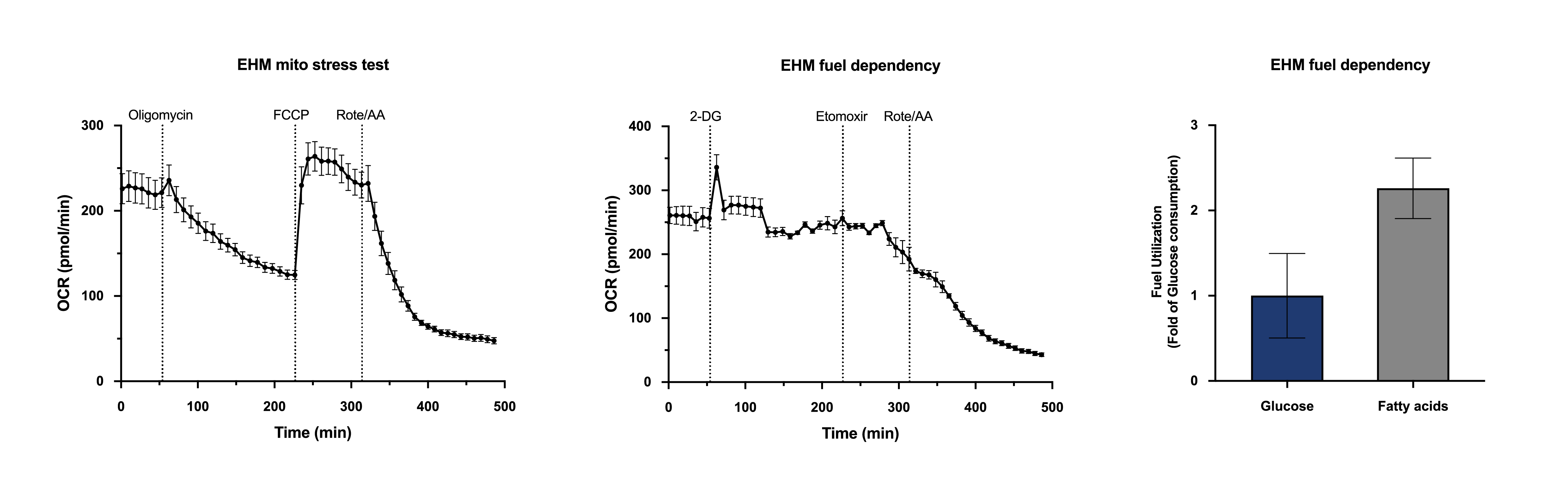
Engineered Heart Muscle (EHM) was evaluated using Seahorse XF metabolic stress and fuel dependency assays (Agilent Technologies) to characterise mitochondrial respiration and substrate use. EHMs display robust oxidative metabolism and metabolic flexibility by using glucose, fatty acids, and alternative substrates for ATP production, reflecting progressive metabolic maturation toward an adult-like cardiac phenotype. EHMs were cultured for 42 days before transferring one EHM per well to a Seahorse XFe24 tissue culture plate. A mitochondrial stress test or fuel dependency test was performed to investigate mitochondrial function and metabolite use. Ventricular Cardiomyocytes were harvested from a 25-day continuous culture before EHM casting.

Representative trace of changes in oxygen consumption rate in EHM when treated with compounds affecting the mitochondrial electron transport chain, shown as mean ± SEM (left).

Representative trace of changes in oxygen consumption rate in EHM when treated with compounds inhibiting processing of glucose (2-deoxy-D-glucose), inhibiting uptake of fatty acids into mitochondria (etomoxir), or inhibiting the electron transport chain, with data shown as mean ± SEM (centre).

Metabolic fuel dependency derived from data in centre shows increased use of fatty acids compared to glucose. Data shown as mean ± SEM (right).

Vial limit exceeded

A maximum number of 20 vials applies. If you would like to order more than 20 vials, please contact us at orders@bit.bio.


Human iPSC-derived ventricular cardiomyocytes

myrCell Ventricular Cardiomyocytes are human iPSC-derived ventricular cardiomyocytes generated using traditional differentiation protocols. Upon revival, the cells exhibit a robust ventricular phenotype characterised by the expression of key cardiac markers including cTnT, α-actinin, NCX1, MYL2, MYH7 and MLC2v.

Ventricular Cardiomyocytes form electrically coupled monolayers and exhibit spontaneous, synchronised contractions within 7 days. They maintain a ventricular-specific electrophysiology, action potential profiles and contractile function for up to 2 months. With organised sarcomeric structures, synchronised beating behaviour and metabolic maturity in both 2D and 3D culture systems, the cells offer a human-relevant cardiac model that bridges the gap between cellular assays and tissue-level physiology.

Ventricular Cardiomyocytes are compatible with high-throughput screening workflows and advanced engineered tissue platforms, making them a versatile tool for both mechanistic studies and translational drug discovery applications. These cells can be used in conjunction with a genetically matched Titin A-band mutation disease model for dilated cardiomyopathy, or paired with Atrial Cardiomyocytes for comparative atrial and ventricular studies.

Benchtop benefits

functional_13

Functional

Cells contract in 2D and 3D, beat spontaneously, exhibit key cardiac markers and ventricular-specific electrophysiology.

Quick-grey_circle

Quick

Ready to use for functional experiments within 7 days post-revival.

3D-Icon

3D compatible

Cells form engineered heart muscle that exhibits progressive maturation and a positive force-frequency relationship

Cells arrive ready to plate

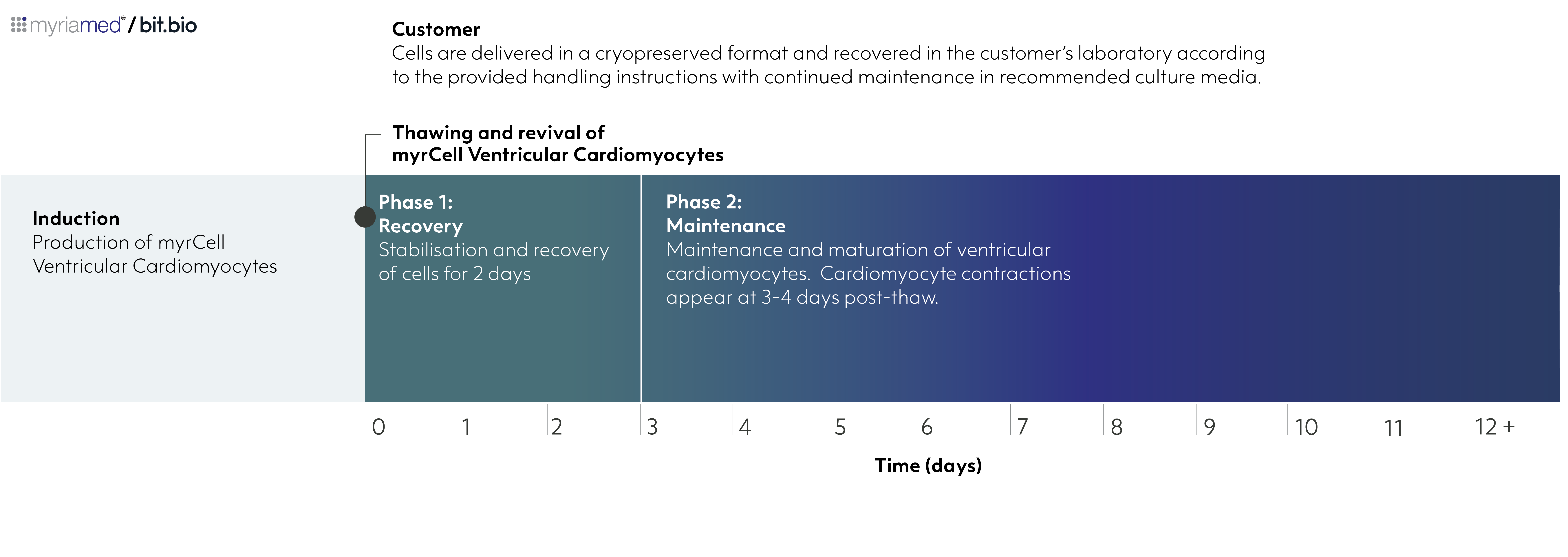
myrcell-ventricular-cardiomyocytes-differentiation-timelineVentricular Cardiomyocytes are delivered in a cryopreserved format and recovered in the customer’s laboratory according to the provided handling instructions with continued maintenance in recommended culture media.

Product specifications

Starting material

Human iPSC line, W001

Karyotype

Normal (46, XY)

Seeding compatibility

6, 12, 24, 48, 96 & 384 well plates

Shipping info

Dry ice

Donor

Male

Vial size

Small: >1 x 10 viable cells

Quality control

Sterility, protein expression (ICC)

Differentiation method

Small molecule-directed differentiation

Recommended minimum seeding density

90,000 cells/cm² (laminin-coated plates); 150,000 cells/cm² (Matrigel-coated plates)

User storage

LN2 or -150°C

Format

Cryopreserved cells

Product use

myrCells are for research use only

Applications

Cardiovascular research
Disease modelling
2D screening and functional assays
3D engineered heart muscle


Scale your study with volume pricing

Enabling scientists to use human cells in their research, running additional experiments without rationing cells or limiting experimental scale


Order quantity Total vials received Pricing tier 
1 - 9 packs 3 - 27 vials Standard price
10 - 33 packs 30 - 99 vials Automatic 10% discount
> 34 packs > 100 vials > Contact us for a quote


Technical data

Mature and spontaneously beating

Ventricular Cardiomyocytes form confluent, electrically coupled monolayers by day 12

myrcell-ventricular-cardiomyocytes-morphology

Brightfield images showing the progressive recovery and attachment of Ventricular Cardiomyocytes cultured on Matrigel-coated flasks. Cells form confluent, electrically coupled monolayers and develop spontaneous, synchronised beating following thawing. Images were acquired at days 1, 4, and 12 post-thaw (10x objective).

Ventricular Cardiomyocytes show spontaneous, synchronised beating by day 7

Representative brightfield video showing spontaneous, synchronised beating of Ventricular Cardiomyocytes cultured in Matrigel-coated flasks at day 7 post-thaw; 4x magnification.

Highly characterised

Ventricular Cardiomyocytes express key cardiac specific markers

myrcell-ventricular-cardiomyoctyes-protein-expression-ctnt-mtcox2

Representative immunofluorescent staining of human iPSC-derived ventricular cardiomyocytes at 10 days post-thaw showing co-localisation of cardiac troponin T (cTnT) with α-actinin, and cTNT with mitochondria (mtCOX2); 60x magnification.

RNA sequencing confirms distinct ventricular and atrial phenotypes

myrcell-cardiomyocytes-rna-seq-ventricular-atrial-phenotypes

RNA sequencing (RNA-seq) demonstrates robust chamber-specific differentiation of Ventricular and Atrial Cardiomyocytes (CM). Differential gene expression and hierarchical clustering reveal distinct ventricular and atrial transcriptomic signatures, confirming reproducible lineage specification and molecular identity across independent differentiation batches. The cells were harvested for RNA-seq at day 7 post-thaw.

Volcano plot showing differential gene expression (A). The x-axis represents log2 fold change (FC) and the y-axis shows -log10 (p value) for each gene. Genes with significant upregulation (blue) or downregulation (red) are labeled, with prominent cardiac-related genes.

Heatmap depicting the relative expression (row Z-score) of selected cardiac-related genes across samples; gene names are listed on the right (B). The heatmap reveals distinct clustering patterns between conditions. The colour scale ranges from -3 (red, indicating lower expression) to +3 (blue, indicating higher expression).

Data were generated from three wild-type iPSC lines (W001, W003, W005) differentiated into both ventricular and atrial cardiomyocytes across 3–4 independent batches. For each line and batch, three technical replicates were pooled prior to sequencing. Ventricular Cardiomyocytes, cat. no. 02-1001-0000, and Atrial Cardiomyocytes, cat. no. 06-1001-0000, are generated from iPSC line W001. Please enquire for further information about iPSC lines W003 and W005.

Technical data

Mature functional phenotype

Robust functional expression of key cardiac ion channels confirmed by automated patch-clamp analysis

myrcell-ventricular-atrial-cardiomyocytes-patch-clamp-ion-channel-activity

Automated patch-clamp analysis confirms robust expression of key cardiac ion channels in Ventricular (red) and Atrial (blue) Cardiomyocytes (CM).

Representative INa current traces with mean ± SEM peak INa amplitude and I-V curve (A). 

Representative ICa,L current traces with mean ± SEM peak ICa,L amplitude and I-V curve fitted with Boltzmann-Ohm curve (B).

Representative trace showing basal inward rectifier current (IK1) in the presence of 20 mM KCl (black) and BaCl2 (grey) and mean ± SEM IK1 at -100 mV (C). 

Measurements were acquired from iPSC-derived cardiomyocytes cultured for 50 days post-thaw. Comparisons were made using unpaired Student’s t-test, Mann Whitney’s U-test vs. Ventricular. ****p<0.0001, *p<0.05; n/N = iPSC-CM/batches.

Mature electrophysiological phenotypes demonstrated by action potential recordings

myrcell-ventricular-atrial-cardiomyocytes-patch-clamp-action-potential

Representative action potential (AP) recordings demonstrate mature ventricular and atrial phenotypes.

Representative membrane voltage (mV) traces showing atrial and ventricular triggered APs in Atrial (blue) and Ventricular (red) Cardiomyocytes (CM) (left).

AP duration at 90% (APD90) and 50% (APD50) repolarisation as mean ±SEM (centre and right).

Measurements were acquired from human iPSC-derived cardiomyocytes cultured for 7 days post-thaw. Comparisons were made using unpaired Student's t-test, Mann Whitney's U-test vs. Ventricular. ****p<0.0001; n = iPSC-CM from one batch.

Chamber-specific contractile properties confirmed by quantitation of contractile dynamics

myrcell-ventricular-atrial-cardiomyocytes-contraction-cytomotion

Contraction analysis using CytoMotion label-free, image-based motion detection software (IonOptix), confirms chamber-specific contractile function of Ventricular (red) and Atrial (blue) Cardiomyocytes (CM).

Frequency (Hz), time to peak (sec), and time to 90% baseline (sec) showed as mean ± SEM (A).

Representative traces showing contraction of human iPSC-derived cardiomyocytes monolayers as mean ± SEM (B).

Measurements were acquired from iPSC-derived cardiomyocytes cultured for 50 days post-thaw. Comparisons were made using unpaired Student's t-test, Mann Whitney's U-test vs. Ventricular. ****p<0.001; n/N = measurement ROI/batches.

Mature mitochondrial function

Mitochondrial stress test confirms metabolically mature bioenergetics

myrcell-ventricular-cardiomyocytes-mitochondrial-function

The Seahorse XF Mito Stress Test demonstrates robust mitochondrial respiration and oxidative metabolism in wild-type Ventricular Cardiomyocytes (WT). Cells were maintained in continuous culture and analysed at two maturation stages (Day 21 and Day 31). Seven days prior to the assay (Day 14 and Day 24), cells were seeded into XFe24 cell culture plates at 80,000 cells per well. On the day of the experiment, mitochondrial function was assessed using the Seahorse XF Mito Stress XF RPMI medium, pH 7.4 supplemented with 10 mM Glucose, 1 mM pyruvate, 200 µM L-AsC, 2% B27.

Representative trace of changes in oxygen consumption rate in Ventricular Cardiomyocytes when treated with compounds affecting the mitochondrial electron transport chain, showed as mean ± SEM (A). 

Mitochondrial respiration, spare respiratory capacity, ATP production and efficiency derived from the representative trace displayed in (A), shown as mean ± SEM (B).

As cardiomyocytes mature from Day 21 to Day 31, they develop enhanced mitochondrial function and bioenergetic capacity, evidenced by increased mitochondrial respiration, higher ATP-linked respiration, and greater spare respiratory capacity. 

Comparisons were made using unpaired Student’s t-test, *p<0.05,  **p<0.01, n = 5.

Functional engineered cardiac tissues

Engineered heart muscle exhibits progressive maturation and a positive force-frequency relationship

myrcell-cardiomyocytes-ehm-protein-expression-mito-actinin

Human iPSC-derived ventricular cardiomyocytes were combined with fibroblasts in a collagen-based hydrogel and cast into myrPlates to generate Engineered Heart Muscle (EHM). Cardiomyocytes were cultured for 7-10 days post thaw before tissue casting. Live video-optical tracking using automated pole detection and peak analysis in the myrImager platform enabled longitudinal assessment of tissue contractility throughout maturation.

Video-optical recording demonstrates spontaneous tissue contraction and enables non-invasive monitoring of functional maturation (upper panel).

Immunofluorescent imaging of the cross section shows a compact, organised tissue with aligned cardiomyocytes, structured sarcomeres and abundant mitochondria (lower panel).

myrcell-ventricular-cardiomyocytes-ehm-force-of-contraction

Video-optical measurements of 16 EHMs over 42 days demonstrated progressive increases in force of contraction (FOC), consistent with continued functional maturation (B). Resting length, quantified as the diastolic pole-to-pole distance in myrPlate cultures, provides a longitudinal readout of tissue compaction and mechanical state. Data are shown as mean ± SEM, n=16.

At study endpoint, EHMs were subjected to isometric organ-bath analysis to independently characterise and validate tissue contractile properties (C). The upper panel shows the preload-dependent Frank–Starling response during 1.5 Hz electrical stimulation at 1.8 mM extracellular Ca2+. Stepwise increases in preload assess the relationship between initial muscle length and force development, a fundamental property of functional myocardium. The lower panel shows the calcium-dependent inotropic response, measured during 1.5 Hz stimulation across increasing extracellular Ca2+ concentrations (0.2–4 mM), providing a measure of maximal calcium-dependent force-generating capacity. Data are shown as mean ± SEM, n=6–12.

Engineered heart muscle exhibits metabolic flexibility and oxidative maturation

myrcell-ventricular-cardiomyocytes-ehm-metabolic-flexibility-oxidative-maturation

Engineered Heart Muscle (EHM) was evaluated using Seahorse XF metabolic stress and fuel dependency assays (Agilent Technologies) to characterise mitochondrial respiration and substrate use. EHMs display robust oxidative metabolism and metabolic flexibility by using glucose, fatty acids, and alternative substrates for ATP production, reflecting progressive metabolic maturation toward an adult-like cardiac phenotype. EHMs were cultured for 42 days before transferring one EHM per well to a Seahorse XFe24 tissue culture plate. A mitochondrial stress test or fuel dependency test was performed to investigate mitochondrial function and metabolite use. Ventricular Cardiomyocytes were harvested from a 25-day continuous culture before EHM casting.

Representative trace of changes in oxygen consumption rate in EHM when treated with compounds affecting the mitochondrial electron transport chain, shown as mean ± SEM (left).

Representative trace of changes in oxygen consumption rate in EHM when treated with compounds inhibiting processing of glucose (2-deoxy-D-glucose), inhibiting uptake of fatty acids into mitochondria (etomoxir), or inhibiting the electron transport chain, with data shown as mean ± SEM (centre).

Metabolic fuel dependency derived from data in centre shows increased use of fatty acids compared to glucose. Data shown as mean ± SEM (right).

Frequently Asked Questions (FAQs)

How long can Ventricular Cardiomyocytes be maintained in culture?

Following thawing and plating at the recommended density, Ventricular Cardiomyocytes establish electrically coupled monolayers and begin spontaneous, synchronised contractions within approximately 7 days. Cells can be maintained in culture for up to 8 weeks, retaining stable electrophysiological properties, ventricular identity, and contractile function.


Do Ventricular Cardiomyocytes beat spontaneously?

Ventricular Cardiomyocytes exhibit spontaneous, synchronised contractions without external stimulation after functional recovery. They are well suited for contractility assays, electrophysiology, calcium handling studies, and safety and toxicity studies.


Can Ventricular Cardiomyocytes be used to generate 3D engineered heart tissues?

Ventricular Cardiomyocytes are optimised for both 2D monolayer cultures and 3D engineered heart muscle (EHM) generation. When combined with cardiac fibroblasts in a collagen-based hydrogel, they form functional tissues that develop physiologically relevant contractile force, tissue elasticity, and mature force-frequency responses.


Are Ventricular Cardiomyocytes suitable for disease modelling?

The wild-type Ventricular Cardiomyocytes serve as the isogenic control for CRISPR-engineered disease models, such as Ventricular Cardiomyocytes TTN A-band mutation, a dilated cardiomyopathy (DCM) disease model. This enables direct comparison between healthy and disease-associated mutations while minimising genetic background variability, making them ideal for mechanistic studies, target validation, and therapeutic testing.


What applications are Ventricular Cardiomyocytes compatible with?

Ventricular Cardiomyocytes are compatible with a broad range of applications, including high-throughput screening, electrophysiology, calcium transient analysis, contractility measurements, cardiotoxicity assessment, transcriptomics, metabolic profiling, and 3D engineered heart tissue studies, providing a versatile platform for cardiovascular drug discovery and translational research.


How are myrCell Ventricular Cardiomyocytes generated?

myrCell Ventricular Cardiomyocytes are derived from the deeply characterised TC1133 human iPSC line using a chemically defined, small molecule-directed differentiation protocol, based on temporal Wnt signalling modulation, followed by metabolic selection to enrich for ventricular cardiomyocytes. Rigorous quality control testing ensures consistent identity and purity across production batches.


ioCells catalogue

Human iPSC-derived cells

powered by opti-ox

Consistent. Defined. Scalable.

bitbio-cell_catalogue_header-with-tracker-Desktop-2500x1664