Human iPSC-derived skeletal myocytes ICC desmin
bit.bio-io6021-skeletal-myocytes-dmd-exon45-deletion-ICC-myocyte-markers
bit.bio-io6021-skeletal-myocytes-dmd-exon45-deletion-morphology
Human iPSC-derived skeletal myocytes ICC desmin
bit.bio-io6021-skeletal-myocytes-dmd-exon45-deletion-ICC-myocyte-markers
bit.bio-io6021-skeletal-myocytes-dmd-exon45-deletion-morphology

cat no | io6021

ioSkeletal Myocytes DMD Exon 45 Deletion

Human iPSC-derived Duchenne muscular dystrophy model

  • Cryopreserved human iPSC-derived cells powered by opti-ox that are ready for experiments in days

  • Study Duchenne muscular dystrophy in a human in vitro model engineered with a DMD exon 45 deletion

  • Consistent, functional model with genetically matched wild type control, suitable for experiments in 2D and 3D muscle bundles

Place your order

per vial

Human iPSC-derived skeletal myocytes ICC desmin

Human iPSC-derived Duchenne muscular dystrophy model

Immunocytochemistry showing robust expression of desmin (red) with DAPI nuclear stain (blue).
bit.bio-io6021-skeletal-myocytes-dmd-exon45-deletion-ICC-myocyte-markers

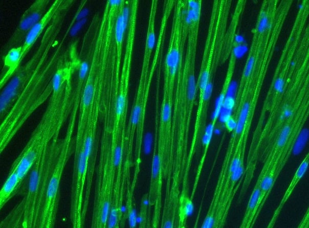
ioSkeletal Myocytes DMD Exon 45 Deletion disease model cells express skeletal muscle cell specific markers and lack expression of dystrophin, demonstrating a Duchenne muscular dystrophy phenotype

Immunocytochemistry staining at day 10 post revival demonstrates robust expression of desmin, a component of the contractile apparatus, and no expression of dystrophin in the ioSkeletal Myocytes DMD Del Ex45/Y disease model cells, whereas ioSkeletal Myocytes, the wild type isogenic control, express both markers (upper panel). Robust expression of myosin heavy chain (MHC) and the muscle transcription factor myogenin is observed in both ioSkeletal Myocytes DMD Del Ex45/Y and ioSkeletal Myocytes (lower panel). Anti-dystrophin antibody clone 2C6 (MANDYS106).

bit.bio-io6021-skeletal-myocytes-dmd-exon45-deletion-morphology

ioSkeletal Myocytes DMD Exon 45 Deletion disease model cells demonstrate classical skeletal myocytes morphology

ioSkeletal Myocytes DMD Exon 45 Deletion form elongated, multinucleated myocytes over 10 days, comparable to the wild-type ioSkeletal Myocytes isogenic control. Day 3 to 10 post-revival; 10X, scale bar 400 um.

Vial limit exceeded

A maximum number of 20 vials applies. If you would like to order more than 20 vials, please contact us at orders@bit.bio.

Human iPSC-derived Duchenne muscular dystrophy model

ioSkeletal Myocytes DMD Exon 45 Deletion are opti‑ox deterministically programmed skeletal myocytes carrying a genetically engineered hemizygous deletion in exon 45 of the DMD gene encoding the dystrophin protein. These cells offer a rapidly maturing, consistent, scalable system to study Duchenne muscular dystrophy in a functional human cell model.

This disease model is part of a Duchenne muscular dystrophy panel of physiologically relevant human iPSC-derived cells that can be incorporated into translational research and drug discovery workflows. The panel includes DMD exon 44, exon 51 and exon 52 deletions. All can be used alongside their genetically matched control ioSkeletal Myocytes.

Benchtop benefits

 
Compare disease model skeletal myocytes with wild type control

Make True Comparisons

Pair the DMD disease model cells with the genetically matched wild type to study the impact of the deletion and to test methods for dystrophin restoration.

Consistent iPSC-derived skeletal myocytes

Consistent

Our platform ensures consistency, scalability, and reproducibility, overcoming the challenges associated with the use of primary muscle cells and immortalised cell lines.

Rapidly maturing skeletal myocytes

Quick

Cells arrive programmed to mature rapidly, forming striated, multinucleated myocytes by day 10 post revival.

Cells arrive ready to plate


bit.bio_ioSkeletal_Myocytes_with_disease_models_timeline

ioSkeletal Myocytes DMD Exon 45 Deletion are delivered in a cryopreserved format and are programmed to mature rapidly upon revival in the recommended medium. The protocol for the generation of these cells is a two-phase process: Phase 1. Stabilisation for 3 days. Phase 2. Maintenance during which the skeletal myocytes mature.

Product specifications

Starting material

Human iPSC line

Karyotype

Normal (46, XY)

Seeding compatibility

6, 12, 24, 48, 96 & 384 well plates

Shipping info

Dry ice

Donor

Caucasian adult male, aged 55-60 years old (skin fibroblast)

Vial size

Large: >5 x 10⁶ viable cells,
Evaluation pack*: 3 large vials of >5 x 10⁶ viable cells

Quality control

Sterility, protein expression (ICC), gene expression (RT-qPCR) and genotype validation (gel electrophoresis)

Differentiation method

opti-ox deterministic cell programming

Recommended seeding density

100,000 cells/cm2

User storage

LN2 or -150°C

Format

Cryopreserved cells

Genetic modification

Hemizygous exon 45 deletion in the DMD gene

Applications

Muscle and neuromuscular research
Disease modelling
Contractility assays
3D muscle tissue engineering

Product use

ioCells are for research use only

* Evaluation packs are intended for first-time users, or for existing users testing a new cell type or derivative. A user can request multiple evaluation packs as long as each one is for a different product.

Technical data

Highly characterised and defined

ioSkeletal Myocytes DMD Exon 45 Deletion disease model cells demonstrate classical skeletal myocytes morphology

iPSC skeletal myocytes DMD exon 45 deletion show classical morphology

ioSkeletal Myocytes DMD Exon 45 Deletion form elongated, multinucleated myocytes over 10 days, comparable to the wild-type ioSkeletal Myocytes genetically matched (isogenic) control. Day 3 to 10 post-revival; 10X objective lens, scale bar 400 um.

Cell culture hacks | human iPSC-derived skeletal myocytes 

Read this blog on skeletal myocytes cell culture for our top tips on careful handling, cell plating and media changes to achieve success from the outset.

bit.bio_ioskeletal myocytes About us

Product resources

(LinkedValues: [{hs_name=ioSkeletal Myocytes , hs_id=161746296924, hs_path=skeletal-myocytes, button_label=View brochure, button_link=https://14527135.fs1.hubspotusercontent-na1.net/hubfs/14527135/Website%20content/Resources/Brochures/bit.bio-ioSkeletal%20Myocytes%20Brochure.pdf, type={label=Brochure, value=Brochure}, thumbnail={alt_text=, width=2550, url=https://14527135.fs1.hubspotusercontent-na1.net/hubfs/14527135/Website%20content/Product%20pages/ioSkeletal%20Myocytes/ioSkeletal-Myocytes-2550px_wide-banner-comp.jpeg, height=1913}, year={label=2024, value=2024}, summary=<p>bit.bio</p>, date_published=1713312000000, sort_date=1713312000000, tags=[{label=ioSkeletal Myocytes, value=ioSkeletal Myocytes}, {label=ioSkeletal Myocytes DMD44, value=ioSkeletal Myocytes DMD44}, {label=ioSkeletal Myocytes DMD52, value=ioSkeletal Myocytes DMD52}, {label=Product information, value=Product information}], media_contact=null, listing_button_label=Download}, {hs_name=Generation of 3D skeletal muscle microtissues using ioSkeletal Myocytes, hs_id=161961134493, hs_path=generation-of-3d-skeletal-muscle-microtissues-using-ioskeletal-myocytes, button_label=View poster, button_link=https://14527135.fs1.hubspotusercontent-na1.net/hubfs/14527135/Product%20collateral/Posters/Stimulating%203D%20Skeletal%20Muscle%20Microtissues%20in%20Perfusable%20Microphysiological%20System_Biond_Poster_MPS_Berlin_Jun23.pdf, type={label=Poster, value=Poster}, thumbnail={alt_text=, width=1932, url=https://14527135.fs1.hubspotusercontent-na1.net/hubfs/14527135/horizontal_ioSkeletal-Myocytes-Muscle-Bundle-Biond-ICC-SAA-Actin%20(1)-1%20(1).png, height=1440}, year={label=2023, value=2023}, summary=<p>Dr Mitchell Han</p> <p>Bi/ond</p> <p>2023</p>, date_published=1710028800000, sort_date=1693267200000, tags=[{label=ioSkeletal Myocytes, value=ioSkeletal Myocytes}, {label=ioSkeletal Myocytes DMD44, value=ioSkeletal Myocytes DMD44}, {label=ioSkeletal Myocytes DMD52, value=ioSkeletal Myocytes DMD52}], media_contact=null, listing_button_label=View }, {hs_name=Human iPSC-derived DMD skeletal myocytes for 3D functional studies and dystrophin restoration, hs_id=173835555607, hs_path=isscr2024-skeletal-myocytes-dmd-3d-function-dystrophin-restoration, button_label=null, button_link=14527135.fs1.hubspotusercontent-na1.net, type={label=Poster, value=Poster}, thumbnail={alt_text=ISSCR24-DMD-Exon44-3D-muscle-bundle (1), width=2953, url=https://14527135.fs1.hubspotusercontent-na1.net/hubfs/14527135/ISSCR24-DMD-Exon44-3D-muscle-bundle%20(1).png, height=1969}, year={label=2024, value=2024}, summary=<p>Bernard, et al</p> <p>bit.bio</p> <p>2024</p>, date_published=1721692800000, sort_date=1721692800000, tags=[{label=ioSkeletal Myocytes, value=ioSkeletal Myocytes}, {label=ioSkeletal Myocytes DMD44, value=ioSkeletal Myocytes DMD44}, {label=ioSkeletal Myocytes DMD52, value=ioSkeletal Myocytes DMD52}], media_contact=<p>Bernard et al,</p> <p>bit.bio</p> <p>2024</p>, listing_button_label=Download}, {hs_name=Introducing ioSkeletal Myocytes | Developing the next generation of human muscle cells, hs_id=161968263500, hs_path=introducing-ioskeletal-myocytes, button_label=null, button_link=null, type={label=Video, value=Video}, thumbnail={alt_text=, width=1318, url=https://14527135.fs1.hubspotusercontent-na1.net/hubfs/14527135/BitBio2022/product-page/Introducing%20ioSkeletal%20Myocytes-min-comp.jpg, height=1000}, year={label=2022, value=2022}, summary=<p><span>Dr Will Bernard | Director of Cell Type Development | </span><span>bit.bio</span></p>, date_published=1707264000000, sort_date=1643068800000, tags=[{label=ioSkeletal Myocytes, value=ioSkeletal Myocytes}, {label=Product information, value=Product information}, {label=ioSkeletal Myocytes DMD44, value=ioSkeletal Myocytes DMD44}, {label=ioSkeletal Myocytes DMD52, value=ioSkeletal Myocytes DMD52}], media_contact=null, listing_button_label=Watch}, {hs_name=Research in Motion with ioSkeletal Myocytes, hs_id=161968263518, hs_path=introducing-human-ipsc-derived-muscle-cells-for-research-and-drug-discovery, button_label=Watch webinar, button_link=https://www.youtube.com/watch?v=TBMR5YNucoMvingin, type={label=Webinar, value=Webinar}, thumbnail={alt_text=, width=738, url=https://14527135.fs1.hubspotusercontent-na1.net/hubfs/14527135/BitBio2022/product-page/ioSkeletal-Myocytes-DDW-ICC-staining-header-image-comp.jpg, height=334}, year={label=2020, value=2020}, summary=<p>Dr Luke Flatt | Senior Scientist | Charles River Laboratories</p> <p>Dr Will Bernard | Senior Scientist | bit.bio</p> <p><span><br><br><br></span></p>, date_published=1707091200000, sort_date=1641340800000, tags=[{label=ioSkeletal Myocytes, value=ioSkeletal Myocytes}, {label=ioSkeletal Myocytes DMD44, value=ioSkeletal Myocytes DMD44}, {label=ioSkeletal Myocytes DMD52, value=ioSkeletal Myocytes DMD52}], media_contact=null, listing_button_label=Watch now}, {hs_name=Advancements in 3D modeling: Building mature, functional 3D skeletal muscle microtissues in vitro, hs_id=161968263527, hs_path=advancements-in-3d-modeling-building-mature-functional-3d-skeletal-muscle-microtissues-in-vitro, button_label=null, button_link=null, type={label=Webinar, value=Webinar}, thumbnail={alt_text=, width=1304, url=https://14527135.fs1.hubspotusercontent-na1.net/hubfs/14527135/Website%20content/Product%20pages/ioSkeletal%20Myocytes/bit.bio_ioskeletal%20myocytes%20About%20us.jpg, height=964}, year={label=2023, value=2023}, summary=<p>Dr Marieke Aarts | Principal Scientist | Bi/ond</p> <p>Amanda Turner | Senior Product Manager | bit.bio</p>, date_published=1710201600000, sort_date=1698710400000, tags=[{label=ioSkeletal Myocytes, value=ioSkeletal Myocytes}, {label=ioSkeletal Myocytes DMD44, value=ioSkeletal Myocytes DMD44}, {label=ioSkeletal Myocytes DMD52, value=ioSkeletal Myocytes DMD52}], media_contact=null, listing_button_label=Watch now}])
Human iPSC-derived DMD skeletal myocytes for 3D functional studies and dystrophin restoration Poster
Human iPSC-derived DMD skeletal myocytes for 3D functional studies and dystrophin restoration

Bernard, et al

bit.bio

2024

Download
ioSkeletal Myocytes Brochure
ioSkeletal Myocytes

bit.bio

Download
Advancements in 3D modeling: Building mature, functional 3D skeletal muscle microtissues in vitro Webinar
Advancements in 3D modeling: Building mature, functional 3D skeletal muscle microtissues in vitro

Dr Marieke Aarts | Principal Scientist | Bi/ond

Amanda Turner | Senior Product Manager | bit.bio

Watch now
Generation of 3D skeletal muscle microtissues using ioSkeletal Myocytes Poster
Generation of 3D skeletal muscle microtissues using ioSkeletal Myocytes

Dr Mitchell Han

Bi/ond

2023

View
Introducing ioSkeletal Myocytes | Developing the next generation of human muscle cells Video
Introducing ioSkeletal Myocytes | Developing the next generation of human muscle cells

Dr Will Bernard | Director of Cell Type Development | bit.bio

Watch
Research in Motion with ioSkeletal Myocytes Webinar
Research in Motion with ioSkeletal Myocytes

Dr Luke Flatt | Senior Scientist | Charles River Laboratories

Dr Will Bernard | Senior Scientist | bit.bio


Watch now

Wild Type and Isogenic Disease Model cells: A true comparison.

Further your disease research by pairing our wild type cells with isogenic disease models.

bitbio-vials-Wild_and_Disease-staggered-2500px_wide

ioCells catalogue

Human iPSC-derived cells

powered by opti-ox

Consistent. Defined. Scalable.

bitbio-cell_catalogue_header-with-tracker-Desktop-2500x1664