14.07.2023 | Published by bit.bio
Human iPSC-derived skeletal myocytes provide a physiologically-relevant tool for research, disease modelling, and high-throughput screening, to study muscle conditions such as Duchenne muscular dystrophy and muscular defects, as well as neuromuscular, and associated metabolic disorders.
To help you get started and achieve the best results when using human iPSC-derived muscle cells we asked Madeleine Garrett, Field Application Specialist at bit.bio, for her top tips for culturing human iPSC-derived ioSkeletal Myocytes and the associated disease models: ioSkeletal Myocytes DMD Exon 44 Deletion and ioSkeletal Myocytes DMD Exon 52 Deletion. Madeleine is a scientist with hands-on experience culturing, optimising and characterising a variety of cell types, in addition to supporting scientists with their technical questions when using bit.bio ioCells.
Compared to other in vitro models, such as C2C12 mouse myoblast cell line or primary muscle cells, ioSkeletal Myocytes have been reported to be fairly straightforward and quicker to culture in addition to offering a consistent, human-relevant system. By following Madeleine’s top tips below, on Geltrex on your cell culture plate, careful handling of cells, frequently and thoroughly re-pipetting cell suspensions, as well as being mindful not to thaw too many vials of cells at once, you should be able to get reliable, consistent results from your experiments every time!
Go straight to the top tips!
- Top tip #1: Control Geltrex temperature to coat the surface of cell culture plates evenly
- Top tip #2: Careful handling of human iPSC-derived skeletal myocytes can improve cell culture success
- Top tip #3: Remix human iPSC-derived skeletal myocytes regularly while in suspension
- Top tip #4: Thaw fewer vials of skeletal myocytes at a time to avoid cell clumping
What are skeletal myocytes?
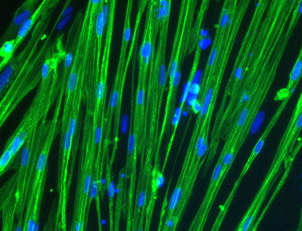
Human skeletal myocytes are one of three types of muscle cells (in addition to smooth and cardiac muscle). They are terminally differentiated muscle cells that represent the smallest building blocks in the larger muscle tissue hierarchy and are critical to a number of physiological functions such as movement and metabolism1. As individual cells, skeletal myocytes express characteristic proteins such as actin, myosin, and dystrophin, all of which are critical for carrying out contractile functions.
Human primary skeletal myocytes are the gold standard for modelling muscle in vitro although they can be limited in supply, lack scalability and each batch needs to be validated due to donor variations, making it difficult to scale for high-throughput screening. Human iPSC-derived skeletal myocytes, such as bit.bio’s precision reprogrammed ioSkeletal Myocytes, are a consistent and scalable alternative to primary muscle cells. In vitro, they form striated, multinucleated myocytes by day 10 post-revival, and cells contract in response to acetylcholine.
"As these cells experience strong, spontaneous contractile forces when in culture, it’s really important to prepare the culture dishes with the right coating for human iPSC-derived skeletal myocytes," advises Madeleine. Here are her top tips!
Top tip #1: Control Geltrex temperature to coat the surface of cell culture plates evenly
In Madeleine’s experience, skeletal myocytes adhere best to cell culture surfaces coated with a Geltrex matrix. Geltrex is a basement membrane extract containing laminin, collagen IV, entactin, and heparin sulphate proteoglycans. She says: “Although Geltrex is fantastic for human iPSC-derived skeletal myocytes, helping them adhere and spread evenly, scientists often don’t realise how quickly it can polymerise when it warms up!” She advises that Geltrex needs to be kept under 4°C, ideally in an icebox, only handling the tube or vial when necessary.
“I suggest keeping your tube or vial on ice the entire time” states Madeleine. “I also advise against freeze-thawing more than 1-2 times, meaning that pre-aliquoting your Geltrex into smaller vials for regular use in the lab can be a lifesaver for cells,” she recommends.
It’s important to prepare a sufficient number of tissue culture plates with a Geltrex coating prior to reviving cryovial(s), aspirating the coating solution from culture plate(s) before seeding your cells. Depending on the volume, smaller aliquots should take about 30 minutes to thaw (from storage at -80°C) at approximately 4°C, and should be kept on ice at all times before coating your cell culture plates.
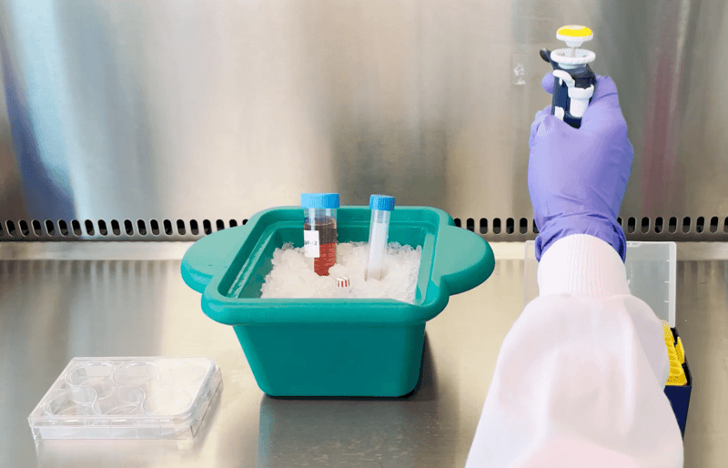 Keeping Geltrex on ice directly before coating plates can result in a more even coating, helping cells adhere and spread out evenly.
Top tip #2: Careful handling of human iPSC-derived skeletal myocytes can improve cell culture success
In the body, skeletal myocytes bundle together to form muscle fibres, therefore it is not surprising that skeletal myocytes can be sticky, meaning that once they attach to a cell culture plate, they adhere extremely firmly. “This has advantages in terms of adherence, and disadvantages in terms of it being difficult to get them off the plate when you need to,” says Madeleine.
Getting the seeding density right! When using ioSkeletal Myocytes, Madeleine recommends a minimum cell seeding density of 100,000 cells/cm2. “If cells are too sparse, they won’t form a monolayer in culture. In fact, in such situations, they will struggle to become established (fuse) and will eventually die. Cells need to be in contact with one another in order to fuse and differentiate into multinucleated skeletal myocytes. If there is an error in seeding (lower seeding density), the cells often look sparse and mononucleated”
For downstream experiments, such as analysis of gene or protein expression, Madeleine advises using a scraping technique with a cell scraper when you lyse and remove myocytes from cell culture plates. "These cells will always stick together, as they're naturally sticky. However, adding the lysis buffer directly to the plates, followed by thorough scraping, is great at helping to reduce stickiness in the resulting cell suspension."
Scientists should always bear in mind that as the human iPSC-derived skeletal myocytes mature in culture, they will start to contract spontaneously, and this contractile function will increase and strengthen with time, meaning that it is normal to witness some cells lifting from the plate. Contraction in the ioSkeletal Myocyte culture can begin as early as 10 days post-revival (see video below).
Madeleine says: “As cells mature, they become stronger, and so do their contractile forces. After a long time (>15 days) in 2D culture, individual cells are even strong enough to come off the plate when they contract. This can even happen with the most perfect Geltrex coating!”.
Short video showing spontaneous contraction of ioSkeletal Myocyte contraction at 0.6 seconds at day 15 post-revival, cells.
Top tip #3: Remix human iPSC-derived skeletal myocytes regularly while in suspension
For best results when thawing and seeding human iPSC-derived skeletal myocytes, such as ioSkeletal Myocytes, it’s important to mix the cells regularly – much more regularly than you might consider doing for other cell types. This is due to the heavy and sticky nature of muscle cells, which means that they can settle and sink out of cell suspensions, and then clump together. This can also result in errors in cell counting impacting the ability to seed cells at the recommended density on cell culture surfaces. Madeleine advises: “Always remember that these cells tend to stick together, and this can significantly affect how they perform in culture. If they are clumped in culture, they have a much higher potential of coming off the plate.”
We recommend periodically pipetting your suspensions thoroughly with a sterile serological pipette (such as a Stripette) to get as close to a single-cell suspension as possible. “A serological pipette works best in my experience, from both a cell handling and ease standpoint,” advises Madeleine. Using a 10 mL serological pipette is useful when thawing ioSkeletal Myocytes, as a more dilute cell suspension will be less likely to clump together. The use of a serological pipette is scalable to larger volumes of cell culture media, and allows you to carry out regular, thorough pipetting of your cell suspensions, making it a useful tool. “The wide diameter of the serological pipette tip also really helps resuspend these cells safely!”
Top tip #4: Thaw fewer vials of skeletal myocytes at a time to avoid cell clumping
The adhesive nature of human iPSC-derived skeletal myocytes means that when handling and plating out cells, maintaining an even cell suspension is critical. “If the cells are left in a vial or tube of media for too long, let’s say if you take out too many cryovials at one time, they’re very likely to adhere together, sink and settle at the bottom. This clumping results in cells that grow poorly on cell culture surfaces and eventually die” states Madeleine. She suggests thawing a maximum of 5 vials at a time if they will be pooled in a larger volume at an early stage or thawing only 2 vials if they will not be pooled. A great way to combat the sticky nature is to work quickly and make sure to thoroughly resuspend the cells at each stage of cell thawing, counting and seeding.
“If you have the odd two or three cells clumped together, you can still get reasonable cell counts and cultures that spread out evenly,” Madeleine continues, “however if you start to get five or more cells regularly clumping together in your cell suspension, the results could be disastrous for your culture and its survival. Some of the cells at the top of these clumps will not be able to attach to the dish, and they’ll eventually die off. This cell death will result in fewer attached cells in the culture, as well as excessive dead cell debris, both of which will negatively impact the health of the culture.”
Summary
In summary, ensure a complete layer of Geltrex on cell culture plates, handle carefully when plating out cell cultures, remember to frequently resuspend cells when they are in a vial or tube, and only thaw one or two vials of frozen cells at a time. Do this, and your cells should be happier and healthier, helping to deliver more reliable results!
Do you have any cell culture tips to share? We’d love to hear from you!
Further details about culturing human iPSC-derived skeletal myocytes can also be found in bit.bio’s ioSkeletal Myocytes User Manual.
About Madeleine Garrett
"Madeleine is a Field Application Specialist at bit.bio. She has significant hands-on experience culturing, optimising and characterising a variety of cell types, in addition to extensive experience in genetic engineering. She routinely supports scientists with their questions about, and experiments using, bit.bio’s human iPSC-derived cells. As part of her role, she is also involved in protocol development and optimisation, cell culture media optimisation and single-cell RNA sequencing. Madeleine has an MSc (preliminary) in Biochemistry and Molecular Biology, a BSc in Biochemistry and Molecular Biology, from Monash University, Melbourne, Australia, and has published in peer-reviewed journals such as EMBO Molecular Medicine."
 Madeleine GarrettField Application Specialist, bit.bio
References
- Willingham TB, Ajayi PT, Glancy B. Subcellular Specialization of Mitochondrial Form and Function in Skeletal Muscle Cells. Front Cell Dev Biol. 2021 Oct 15;9:757305. doi: 10.3389/fcell.2021.757305.