.png?width=1715&height=651&name=ioGlutamatergic_neurons-PRKN-R275W-R275W-Morphology%20(1).png)


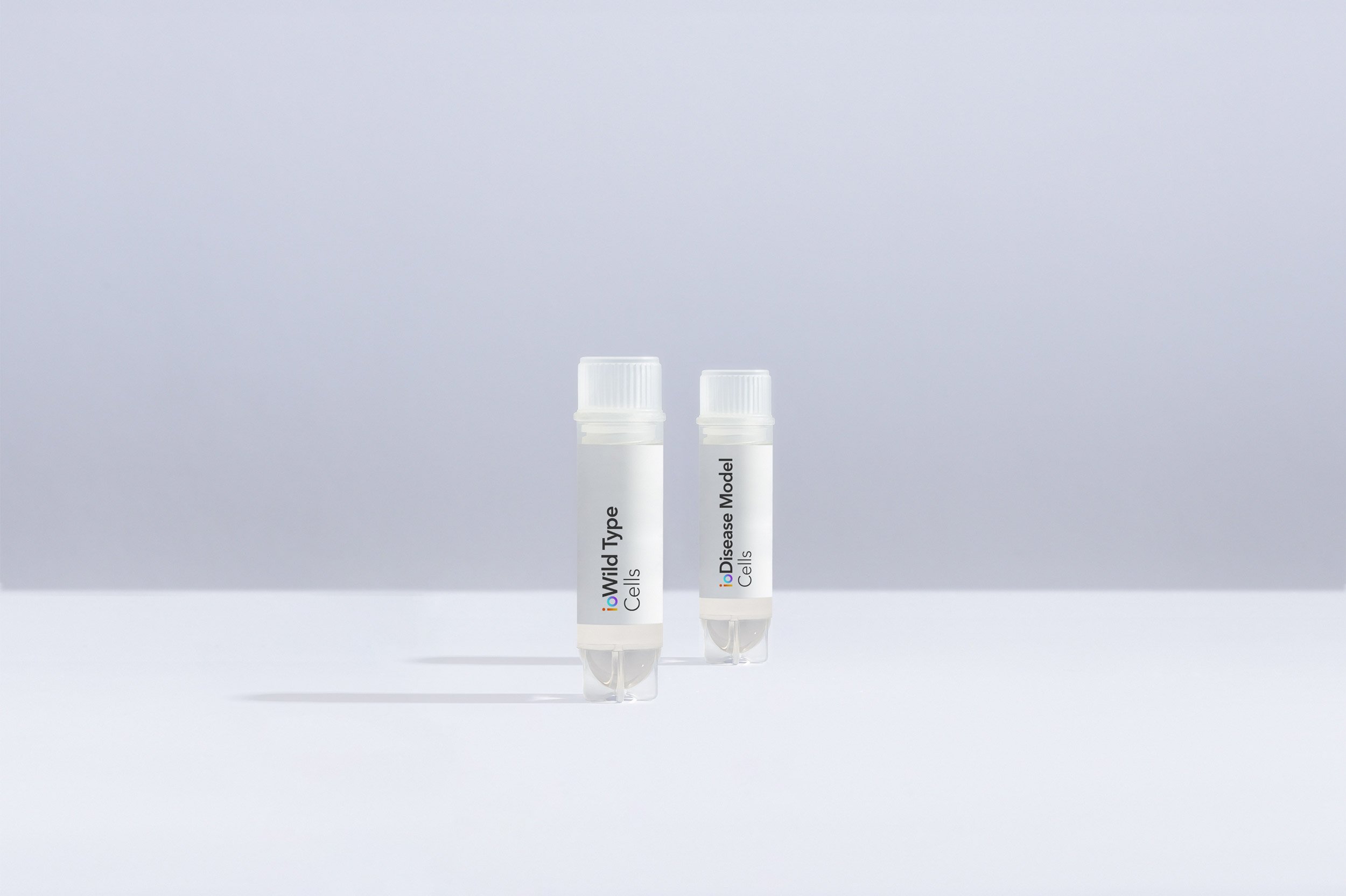
cat no | io1020
ioGlutamatergic Neurons PRKN R275W/R275W
Human iPSC-derived Parkinson's disease model
-
Cryopreserved human iPSC-derived cells powered by opti-ox that are ready for experiments in days
-
In vitro cell model engineered to carry a mutation in PRKN for Parkinson's disease research
-
Consistent, functional excitatory neurons that form neuronal networks within days
Human iPSC-derived Parkinson's disease model
ioGlutamatergic Neurons Media Kit
Cell culture media kit for the culture of ioGlutamatergic Neurons up to 14 days post-thaw
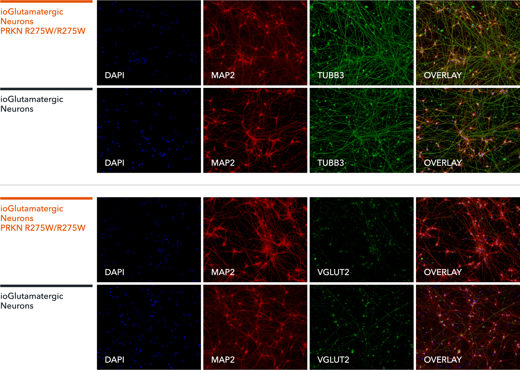
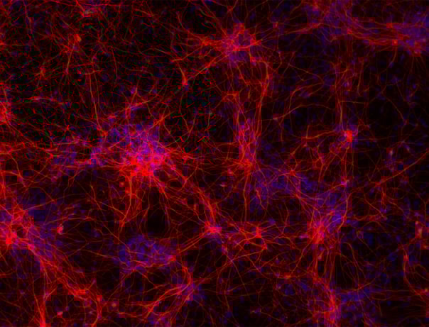
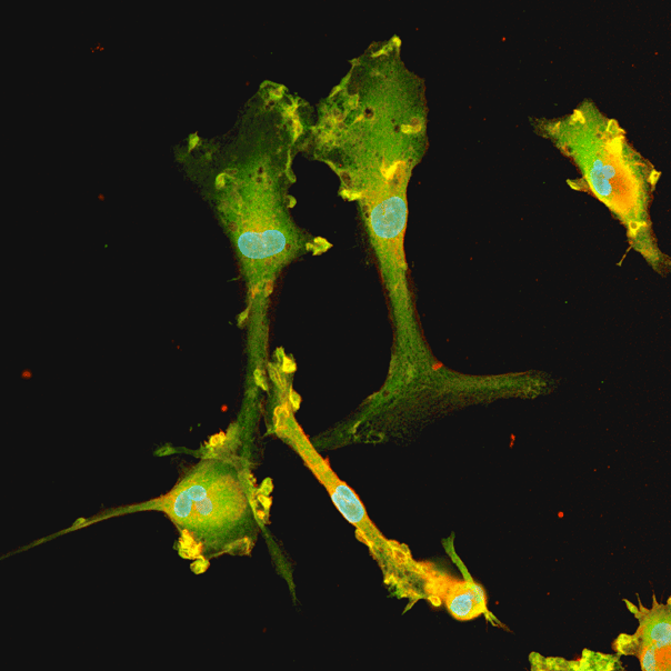
ioGlutamatergic Neurons PRKN R275W/R275W express neuron-specific markers comparably to the wild-type control
Immunofluorescent staining on post-revival day 11 demonstrates similar homogenous expression of pan-neuronal proteins TUBB3 and MAP2 (upper panel) and glutamatergic neuron-specific transporter VGLUT2 (lower panel) in ioGlutamatergic Neurons PRKN R275W/R275W compared to the genetically matched control. 100X magnification.
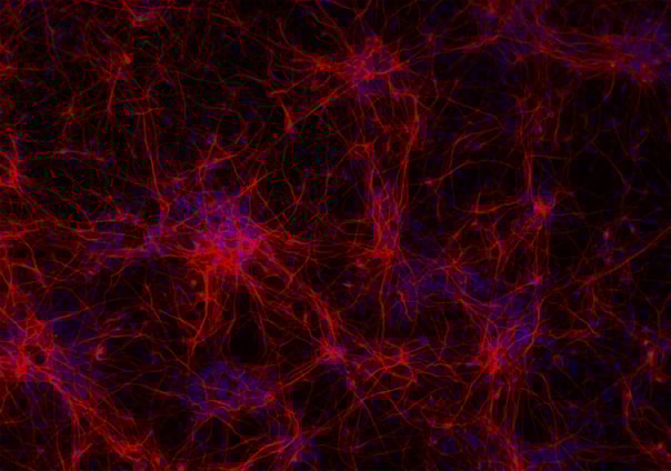
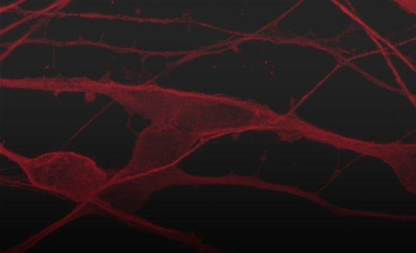
ioGlutamatergic Neurons PRKN R275W/R275W form structural neuronal networks by day 11
ioGlutamatergic Neurons PRKN R275W/R275W mature rapidly, show glutamatergic neuron morphology and form structural neuronal networks over 11 days, when compared to the wild-type control. Day 1 to 11 post thawing; 100X magnification.
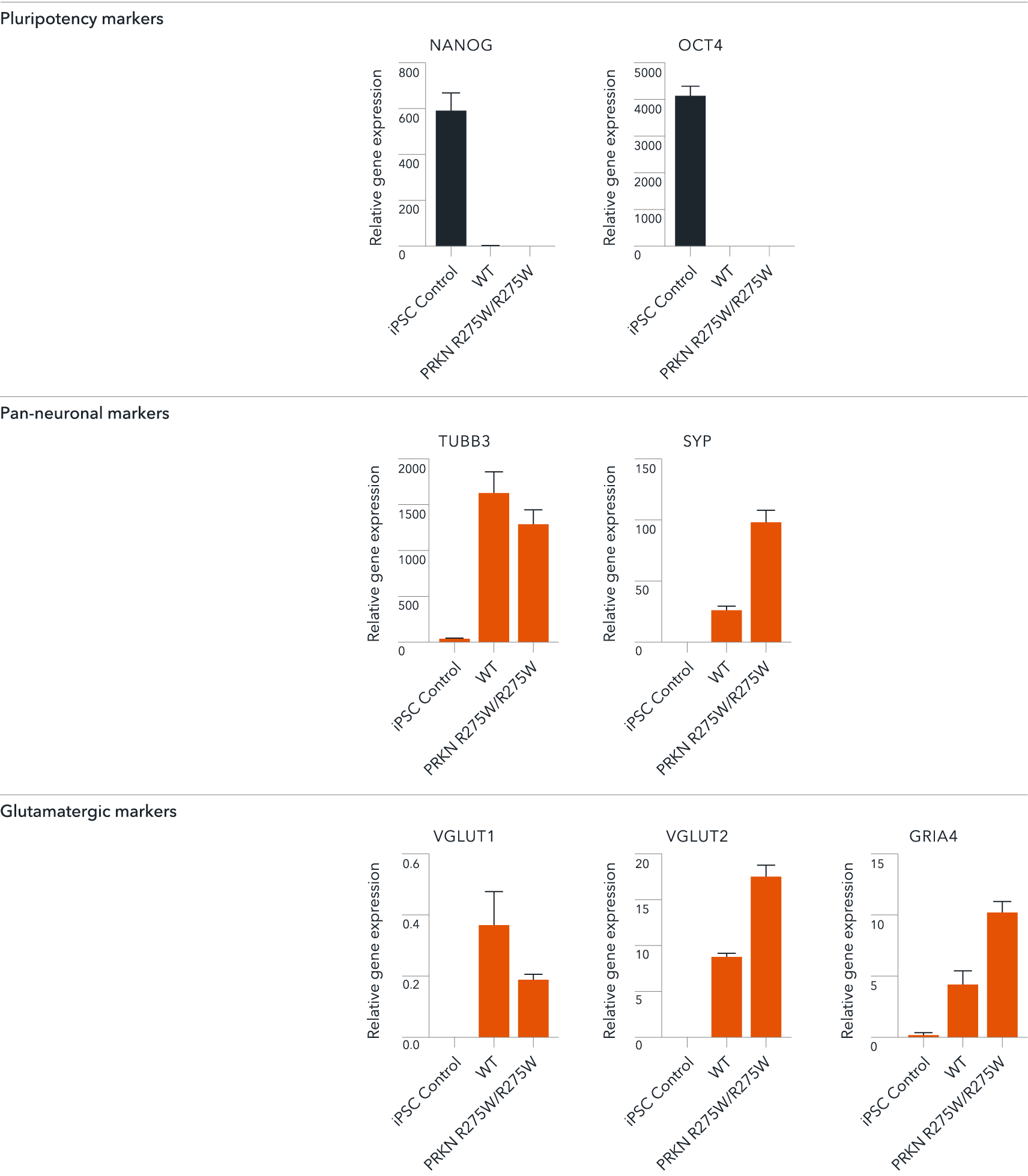
ioGlutamatergic Neurons PRKN R275W/R275W demonstrate gene expression of neuronal and glutamatergic-specific markers following deterministic programming
Gene expression analysis demonstrates that ioGlutamatergic Neurons PRKN R275W/R275W and the wild-type control (WT) lack the expression of pluripotency markers (NANOG and OCT4) at day 11, whilst robustly expressing pan-neuronal (TUBB3 and SYP) and glutamatergic specific (VGLUT1 and VGLUT2) markers, as well as the glutamate receptor GRIA4. Gene expression levels were assessed by RT-qPCR (data normalised to HMBS; cDNA samples of the parental human iPSC line (hiPSC) were included as reference). Data represents day 11 post-revival samples, n=2 replicates.
View the step-by-step RNA extraction and RT-qPCR protocol used to generate this data
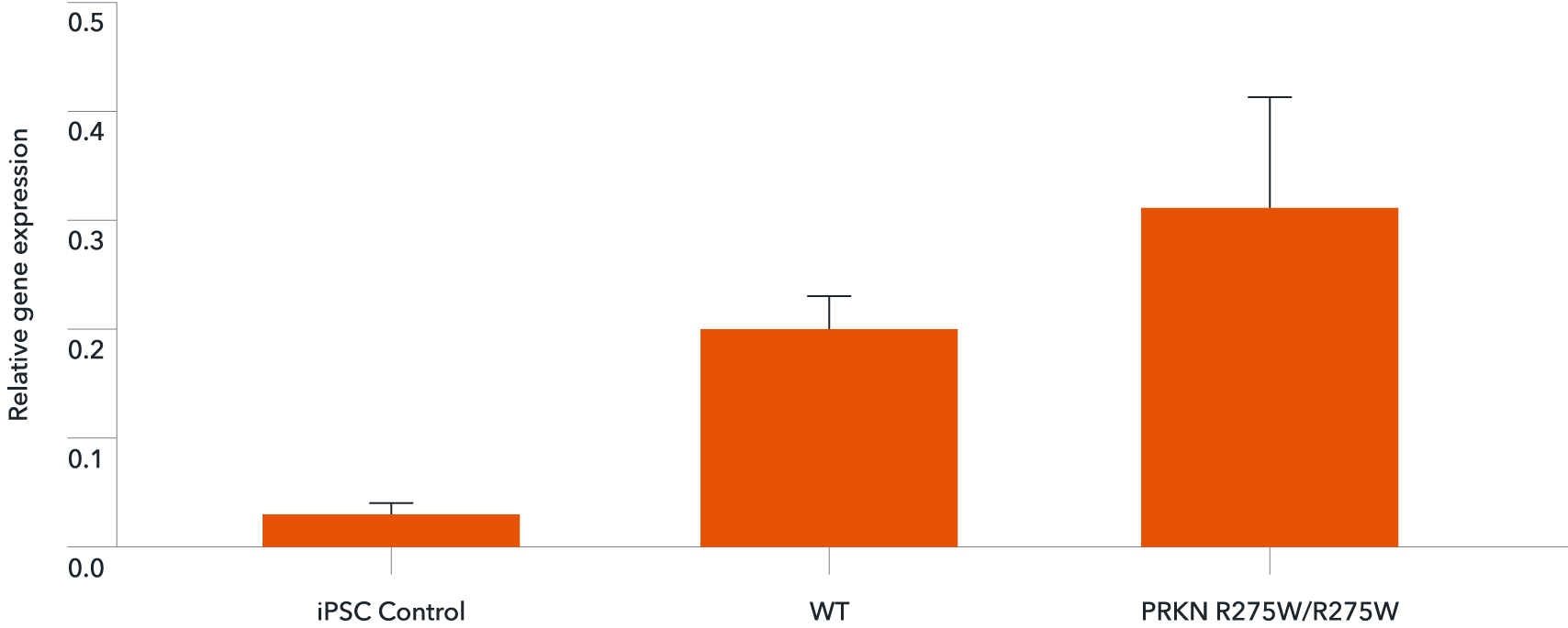
Disease-related PRKN is expressed in ioGlutamatergic Neurons PRKN R275W/R275W following deterministic programming
RT-qPCR analysis demonstrates expression of the PRKN gene in both wild type ioGlutamatergic Neurons (WT) and ioGlutamatergic Neurons PRKN R275W/R275W at day 11 post revival. Data normalised to HMBS, n=2 replicates.
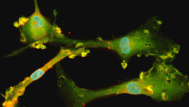
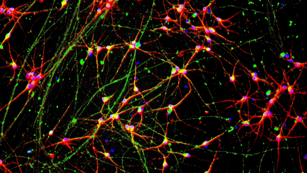
Phenotypic characterisation of a human iPSC-derived tri-culture using ioGlutamatergic Neurons, ioAstrocytes, and ioMicroglia
Using our fully optimised protocol, ioGlutamatergic Neurons (MAP2, red), ioMicroglia (IBA1, yellow) and ioAstrocytes (vimentin, cyan) were co-cultured to create a highly defined CNS model. High-resolution ICC analysis confirms the successful co-localisation and morphological health of three distinct cell types within a unified environment. By day 7, the protocol yields a highly consistent, integrated network suitable for complex cell modelling. DAPI (blue) highlights the total cell density and integrity of the culture. This protocol is compatible with derivative products of the three cell types, ensuring straightforward implementation across experimental workflows.
Efficient mRNA transfection into ioGlutamatergic Neurons
ioGlutamatergic Neurons are efficiently transfected and show sustained long-term expression of mRNA encoding GFP. ioGlutamatergic Neurons were imaged from day 1 post-thaw and throughout the experiment to assess transfection efficiency and evaluate potential cytotoxic effects of the transfection protocol. Day 1 images were captured prior to transfection on the same day.
Download the step-by-step protocol for lipid-based delivery of synthetic mRNA into ioGlutamatergic Neurons.
Lipid-based delivery of synthetic mRNA into ioGlutamatergic Neurons
ioGlutamatergic Neurons were transfected 24 hours post-thaw using Lipofectamine™ Stem Transfection Reagent. The transfection efficiency was evaluated by fluorescence imaging over 18 days after mRNA delivery, resulting in high transfection efficiency (close to 100%) and long-term sustained GFP expression.
Quantification of the GFP signal shows a decrease in GFP intensity over time, while the percentage of GFP-positive cells remains largely unchanged over time.
(A) The percentage of GFP-positive cells from two independent experiments.
(B) GFP intensity, quantified in successfully transfected cells from two independent experiments is quantified and normalised to day 2 (24 hours post-transfection).
Industry leading seeding density
The recommended minimum seeding density is 30,000 cells/cm2, compared to up to 250,000 cells/cm2 for other similar products on the market. One small vial can plate a minimum of 0.7 x 24-well plate, 1 x 96-well plate, or 1.5 x 384-well plates. This means every vial goes further, enabling more experimental conditions and more repeats, resulting in more confidence in the data.
Vial limit exceeded
A maximum number of 20 vials applies. If you would like to order more than 20 vials, please contact us at orders@bit.bio.

.png?width=1715&height=651&name=ioGlutamatergic_neurons-PRKN-R275W-R275W-Morphology%20(1).png)


_MAP2(R)_DAPI(B)_%20(1).png?width=604&name=a-HTT50CAGWT_Overlay__TUBB3(G)_MAP2(R)_DAPI(B)_%20(1).png)


Hoescht(blue)TUBB3(blue)_day4.png?width=604&name=bit.bio_ioGlutamatergic%20Neurons_60xMAP2(red)Hoescht(blue)TUBB3(blue)_day4.png)


.png?width=604&name=a.HTT50CAGWT__TUBB3(G).png)


-1.png?width=604&name=CRL%20video%20%231%20card%20for%20webpage%20(thinner%20gradient%20line)-1.png)


Hoescht(blue)TUBB3(blue)_day4.jpg?width=604&name=bit.bio_ioGlutamatergic%20Neurons_60xMAP2(red)Hoescht(blue)TUBB3(blue)_day4.jpg)

.png?width=1860&height=1260&name=bit.bio_3x2_ioGlutamatergic%20Neurons_MAP2_Hoescht_x20_hi.res%20(1).png)