03.08.2026 | Published by bit.bio
Accurate cell counting is the foundation of a successful experiment. For human iPSC-derived cells, which are more sensitive than immortalised cell lines, getting the count right is critical for ensuring correct seeding densities and long-term culture health.
In our recent webinar, "Cell counting workshop | Top tips for human iPSC-derived cells," bit.bio experts Charlotte Durham, BSc (Operational Quality Manager) and Jack Gowen, MSc (Senior Scientist, QC and GMP Analytical Development) shared their internal benchmarking data and bench-side best practices to help you master your workflow.
Whether you are mastering how to use a hemocytometer for the first time or fine-tuning your automated workflow, these six tips will help you achieve more reliable, reproducible cell counting results.
Go straight to the top tips!
- Top tip #1: Choosing your counting method
- Top tip #2: Mind the clock when using viability stains
- Top tip #3: Standardise your thawing and resuspension
- Top tip #4: Check your equipment and loading technique
- Top tip #5: Use proper handling to prevent "clumping" errors
- Top tip #6: Mitigate edge bias and perform replicate counts
Top tip #1: Choosing your counting method
The first step is deciding between manual and automated counting. "One of the great benefits of automated cell counters is the removal of the human element, promoting greater standardisation across users and labs," notes Charlotte Durham.
However, a manual hemocytometer remains an invaluable and widely accessible tool for lab users due to its ease of use and low cost. As Charlotte explains, "As long as best practices are followed, this method is perfectly fine for cell counting. There is a reason it's still used." Whether you use an automated instrument or a traditional hemocytometer, being consistent in your approach is the most critical factor for success.
Top tip #2: Mind the clock when using viability stains
The two most common methods for assessing cell viability are Trypan Blue and fluorescent stains like Acridine Orange/Propidium Iodide (AO/PI). While Trypan Blue is the gold standard when using a hemocytometer, it requires careful timing.
"Trypan blue can actually be toxic to cells over time, so if counting is taking longer, reduced viability may be observed that is not actually reflective of cell health at the time," warns Charlotte. If your cell counting process is taking too long, automated counters that utilise fluorescent dyes like AO/PI or DAPI can be highly beneficial. Ultimately, "using any viability stain will help increase the consistency of your counts."
Top tip #3: Standardise your thawing and resuspension
Proper cell counting actually starts the moment you pull your vial from the freezer. "Cryopreserved cells are delicate at thaw, and it's important to remember that they are within a cryopreservant themselves," Jack advises. He recommends thawing them strictly in a 37°C water bath for around two minutes to prevent prolonged toxic exposure.
Furthermore, Jack stresses the importance of resuspending your cells in the correct basal media rather than generic lab buffers: "Read the user manual to ensure that you are resuspending them properly in the correct media, because other buffers such as DPBS may have a negative impact on your cells or the counting method of choice."
Top tip #4: Check your equipment and loading technique
Before capturing any data, ensure your equipment is set up to succeed. If you are learning how to use a hemocytometer, Jack recommends checking your slide-loading technique: "Ensure that the cover slip is properly applied and the Newton's rings are present, so you should see a small rainbow effect on and through the cover slip."
For automated systems, ensure the internal microscope is perfectly focused and free of air bubbles. Any bubbles in the viewing chamber will prevent the cells from forming a full field of view, drastically reducing the statistical significance of your cell counting results.
Top tip #5: Use proper handling to prevent "clumping" errors
Human iPSC-derived cells, particularly neurons, can be "sticky" and prone to clustering (appearing as "grape-like" clusters), which will throw off your numbers regardless of the method you choose.
To create a homogenous suspension, "make sure you are pipetting the sample gently and avoid vigorous pipetting or even vortexing the cells, because this will again damage the cell health," explains Jack. To avoid variations caused by cells settling at the bottom of the vial before they reach your hemocytometer, Jack notes: "It's important you don't let the cells settle and that when you are sampling from the middle of the tube."
Top tip #6: Mitigate edge bias and perform replicate counts
When performing manual cell counting, it is essential to count enough squares and avoid double-counting cells. Jack advises counting the four outer corner squares of the hemocytometer (Figure 1. B, C, D & E) to achieve a statistically significant count of around 50 to 100 cells.
To avoid "edge bias", establish a strict rule for cells resting on the grid lines: "We recommend counting either the left and top line of the square, or you are free to count the bottom and right side of the square. That's to make sure you're not double counting cells."
Finally, always calculate your statistical variance. "We recommend and follow a practice here at bit.bio of making sure that [replicate counts] are below 20% CV [coefficient of variation]," says Jack. If your variance between counts is higher than 20%, you should omit statistical outliers, gently re-mix your sample, and perform a recount to ensure robust data.
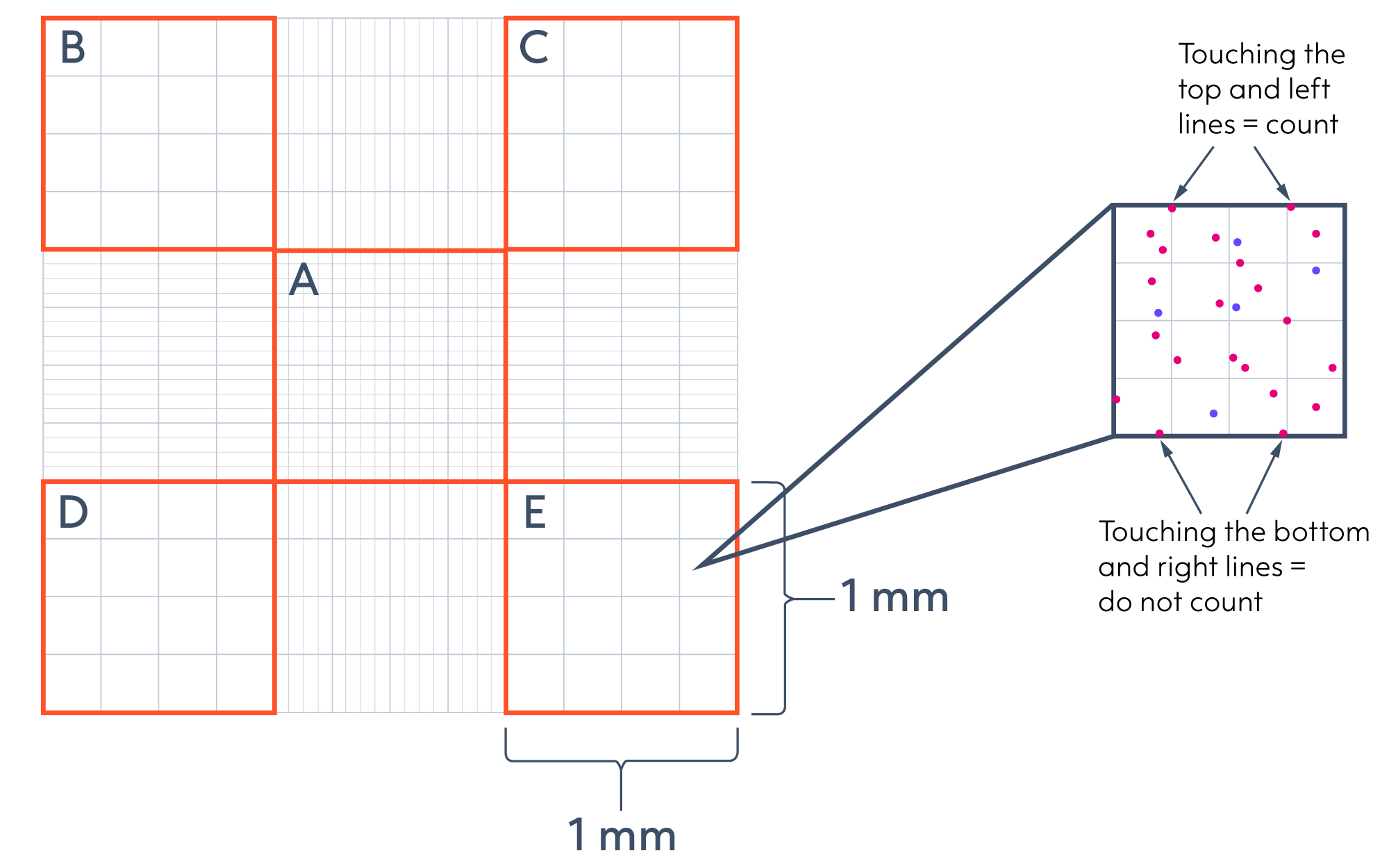
Figure 1. Schematic diagram of a hemocytometer. Recommendations for rules to follow when counting cells.
Master your cell counting workflow
By implementing these small but significant changes, you can significantly reduce the variability in your experiments and ensure that every vial of ioCells you thaw performs to its full potential.
Ready to dive deeper?
Watch the full Cell Counting Workshop webinar to see the benchmarking data and live demonstrations from Jack and Charlotte.
Ready to implement these tips at the bench?
Download our cell counting protocol for ioCells
Explore the bit.bio resource hub for more top tips
FAQs
1. When learning how to use a hemocytometer, how do I avoid double-counting cells?
To ensure accuracy and prevent "edge bias" when using a hemocytometer, follow a strict boundary rule for any cells that land directly on the grid lines. While standard protocols can vary between different institutions, bit.bio expert Jack Gowen recommends focusing on the top and left lines:
-
Count any cells touching the top and left boundary lines of the square
-
Ignore any cells touching the bottom and right lines
Sticking to this specific "L-shape" border configuration ensures that cells resting on shared boundaries are only counted once. This eliminates edge bias and prevents you from accidentally inflating your total cell counting results.
2. How many cells should I aim to count for an accurate result?
For the most reproducible results, you should aim for a concentration within the linear range of your counting method. When using a hemocytometer, aim to count roughly 50 to 100 cells across your chosen squares (typically the four outer corners). If your sample is too concentrated, you increase the risk of clumping. If it is too dilute, the statistical variance, measured as your %CV, will exceed the recommended 20% limit, leading to inconsistent seeding densities in your downstream assays.
About Charlotte Durham
Charlotte Durham, BSc, is an Operational Quality Manager with over ten years of experience within the biotechnology industry. Currently leading cross-functional initiatives at bit.bio, Charlotte possesses dual expertise in Quality Assurance and Quality Control, specialising in designing scalable processes that drive efficiency and continuous improvement. Notably, she established bit.bio’s QC Department for the release testing of iPSC-derived products, and she continues to oversee in-house laboratory operations, assay validation, and risk-based batch dispositions.
 Charlotte Durham, BSc
Operational Quality Manager at bit.bio
About Jack Gowen
Jack Gowen, MSc, is a Senior Scientist with over seven years of experience in quality control within the UK pharmaceutical and biotech sectors. Currently specialising in QC and Analytical Development at bit.bio, Jack brings deep expertise to assay development and cell-based assays utilising human iPSC-derived cells. He holds an MSc in Biotechnology from the University of Nottingham.
 Jack Gowen, MScSenior Scientist QC and GMP, Analytical Development at bit.bio