Custom ioDisease Model Cells™


Your mutation of interest in our opti-ox™ powered human iPSC-derived cells

CDM Image 2-1


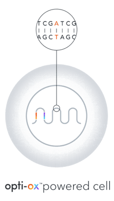
With this custom offering, you can now order your mutation of choice in bit.bio’s opti-ox™ powered human-iPSC derived cells and receive consistent, defined, cryopreserved disease models that can be immediately incorporated into your experiments.

Every custom disease model comes with a genetically matched control, ioWild Type Cells™, giving you confidence that even subtle variances in your data are attributable to your mutation of interest.

Remove the challenges associated with animal models, patient-derived cells and directed differentiation protocols from your workflows, and start collecting disease-relevant data you can trust in a human context. Each project begins as a conversation with our experts about your custom disease model needs.

Pair with a genetically matched control

Build your custom disease model into these wild type cell backgrounds. Use the genetically matched control in your experiments to make true comparisons in your data, and confidently link genotype to phenotype. 

ioGlutamatergic Neurons™ ioWild Type Cells
ioGlutamatergic Neurons™ cat no. io1001
Learn more
ioSkeletal Myocytes™ ioWild Type Cells
ioSkeletal Myocytes™ cat no. io1002
Learn more
ioMicroglia™ | Male ioWild Type Cells
ioMicroglia™ | Male cat no. io1021
Learn more
ioGABAergic Neurons™ ioWild Type Cells
ioGABAergic Neurons™ cat no. io1003
Learn more

Insights from our partners on Custom ioDisease Model Cells

"The access to better human cell-based in vitro disease models as generated within our partnership is transformational for the way we do drug discovery with our pharma and biotech clients. We can now provide discovery studies in human stem cell-based assays, where previously we would only do this in for example immortalised cell lines or more simple assay types.

Marijn Vleming


      Marijn Vlaming, PhD     
      Charles River Laboratories


Uncover subtle phenotypes

Identifying intrinsic, observable phenotypes in ioDisease Model Cells has the potential to streamline compound testing in drug discovery by reducing the need to induce disease-related phenotypes when modelling specific diseases. Discover the data generated by our industry partners below.

Data on our
disease models
Data on our
disease models

Huntington's disease

A Huntington's disease-relevant phenotype has been observed in ioDisease Model Cells devleoped for industry partners.
uncover subtle phenotypes in HD 2
In this example, Charles River Laboratories carried out functional characterisation of ioGlutamatergic Neurons HTT 50CAG/WT by using MaxWell’s MaxTwo high-density microelectrode array (MEA) platform. The data demonstrates a significant decrease in firing rate compared to wild-type isogenic control.
Now, Charles River Laboratories use this disease model in their End-to-End Huntington’s Disease Studies offering. Their customers can access in vitro assays using a disease model that recapitulates the Huntington’s disease phenotype. 

Frontotemporal dementia (FTD)

Our disease models of tau dysfunction show a tau hyperphosphorylation phenotype important to frontotemporal dementia research.

uncover subtle phenotypes in FTD_ALS 1

A panel of disease models for studying frontotemporal dementia (FTD) was developed by engineering either FTD-causing heterozygous or homozygous N279K or P301S mutations in the MAPT gene of ioGlutamatergic Neurons.

At day 21 post-revival, the data showed hyperphosphorylation of mutant tau protein compared to the wild type control using high-content image analysis. The panel offers the potential to investigate the impact of mutant tau on the molecular mechanisms of disease and test potential drug candidates rapidly in high throughput multiwell plate format. Data courtesy of Charles River Laboratories.


Amyotrophic lateral sclerosis (ALS)

ioDisease Model Cells built to study ALS show a significantly reduced neuronal firing phenotype compared to the isogenic control. 
uncover subtle phenotypes in ALS 4 copy

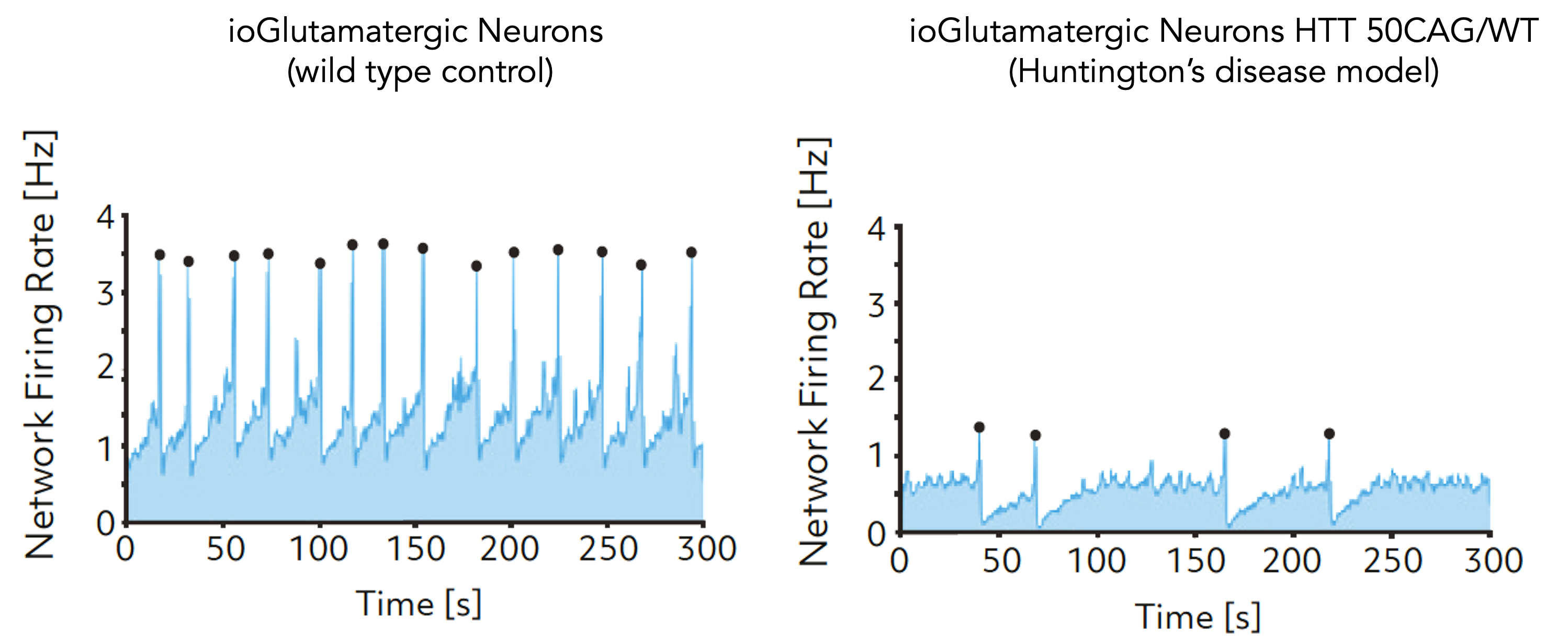
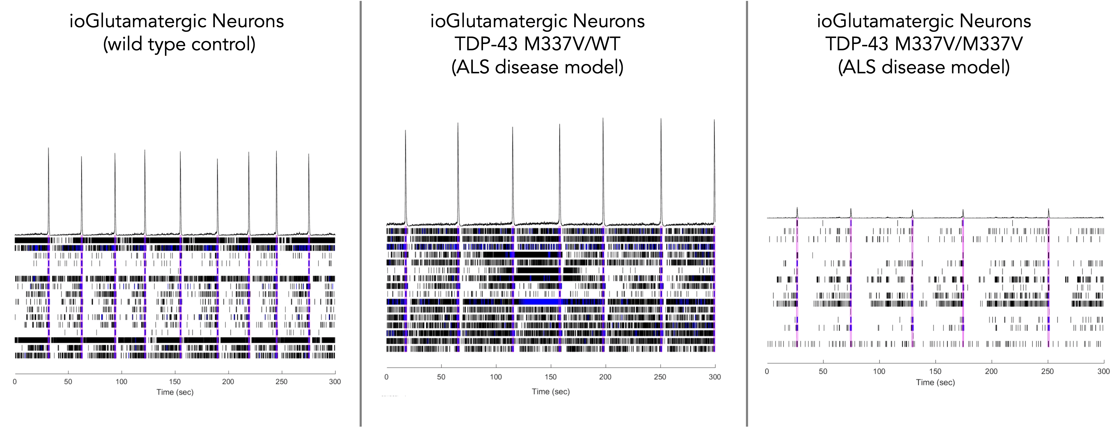
Using a microelectrode array (MEA)-based assay our ALS disease model (ioGlutamatergic Neurons TDP-43 M337V/M337V™) demonstrates a significant reduction in neuronal activity compared to the ALS disease model with a heterozygous mutation (ioGlutamatergic Neurons TDP-43 M337V/WT™) and the wild-type isogenic control. Data courtesy of Charles River Laboratories. 

This indicates their potential as a relevant translational in vitro drug discovery model for ALS.

Charles River Laboratories Discovery team now offers these disease models alongside high-content imaging and gene expression analysis to support investigations into TDP-43 protein phosphorylation, mislocalisation, aggregation, and altered mRNA splicing, which are hallmarks of ALS and FTD. 

Duchenne muscular dystrophy (DMD)

ASO-mediated Dystrophin restoration  demonstrated in ioSkeletal Myocytes DMD Exon 44 Deletion disease model cells
ioSkeletal Myocytes DMD Exon 44 Deletion disease model cells show dystrophin restoration by ASO-mediated exon skipping

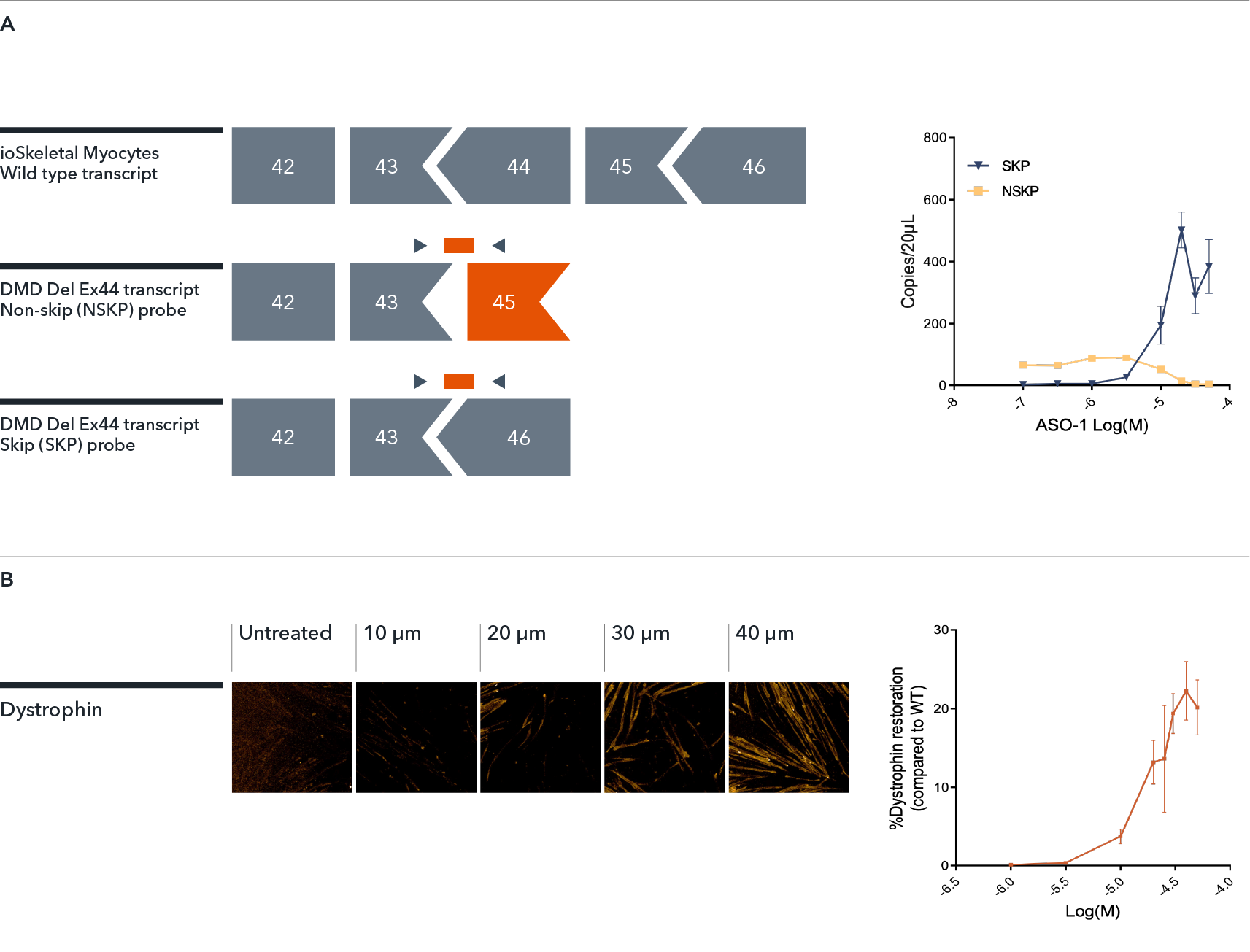
The DMD disease model offers a valuable translational model to investigate methods for dystrophin restoration, such as ASO-mediated exon skipping.

In this example, ioSkeletal Myocytes (wild type control) and ioSkeletal Myocytes DMD Exon 44 Deletion (DMD Del Ex44) were treated by gymnosis with exon 45 skipping antisense oligonucleotide (ASO-1). The data shows a concentration-dependent increase in the amount of mRNA transcript (A) and dystrophin protein (B) in the DMD exon 44 deletion disease model cells, indicating that ASO-1 treatment has been successful in creating an in frame mRNA transcript for dystrophin that leads to protein expression. Data courtesy of Charles River Laboratories.

Interested in generating your own ioDisease Model Cells?

Start a no-commitment project consultation with our experts today.

Bit.bio_office_Pic0053compressed

Related pages

Discover ioCells Learn about our range of human iPSC-derived cells for research and drug discovery
Our platform Discover the cell identity coding platform behind our cells
About us Find out more about us and our people