


cat no | io1001
ioGlutamatergic Neurons
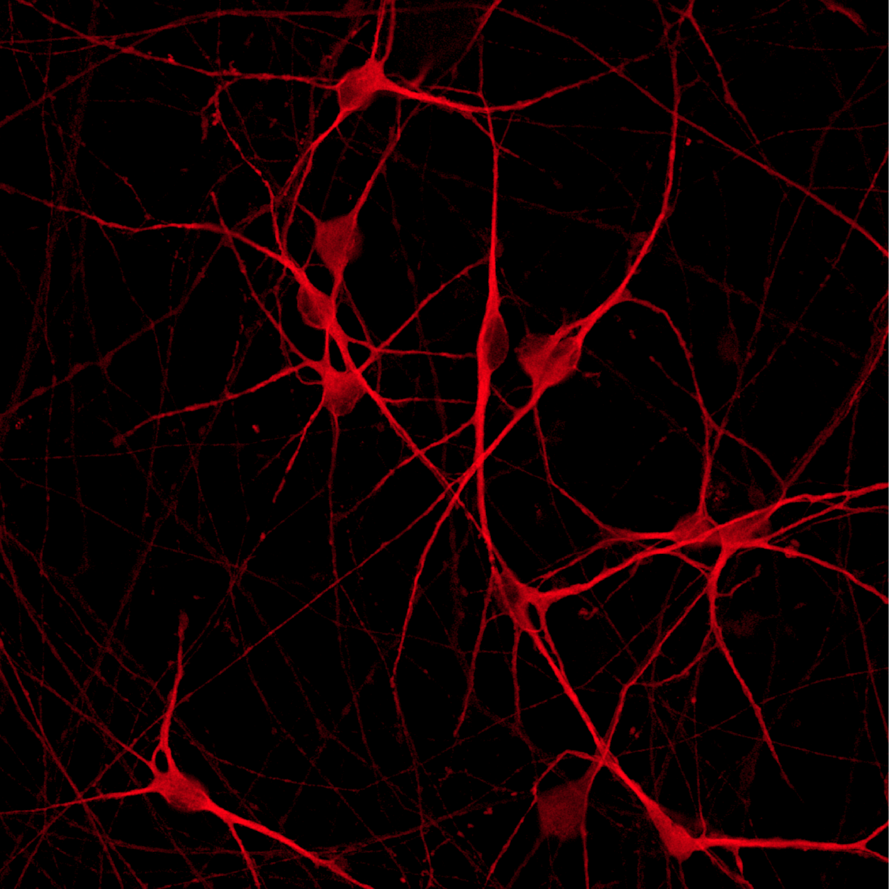
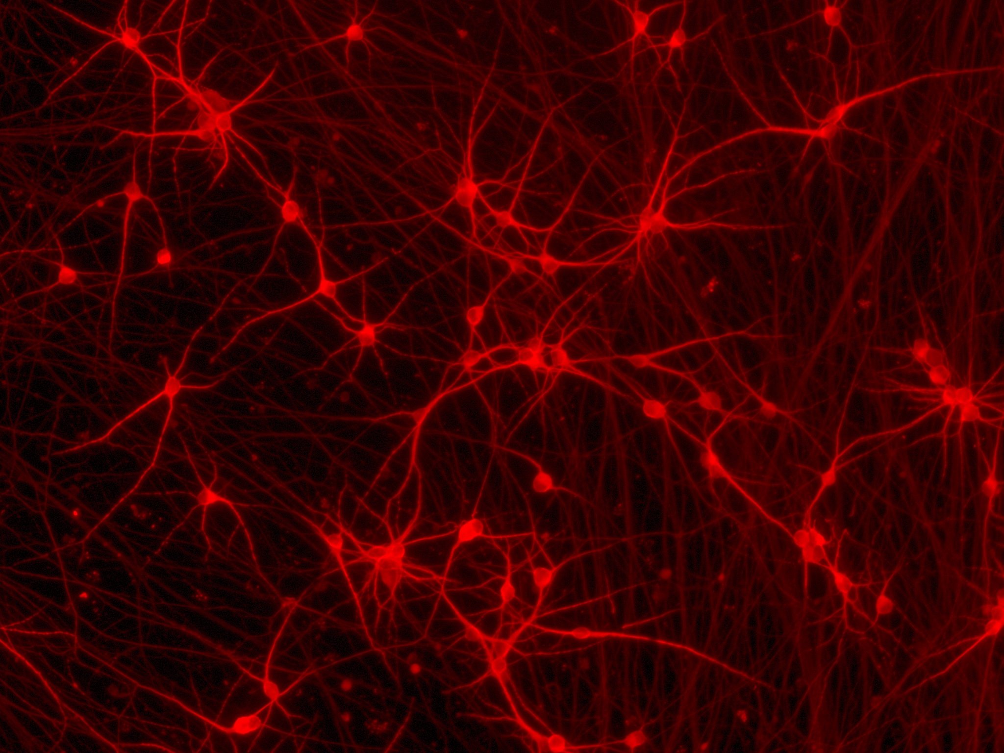
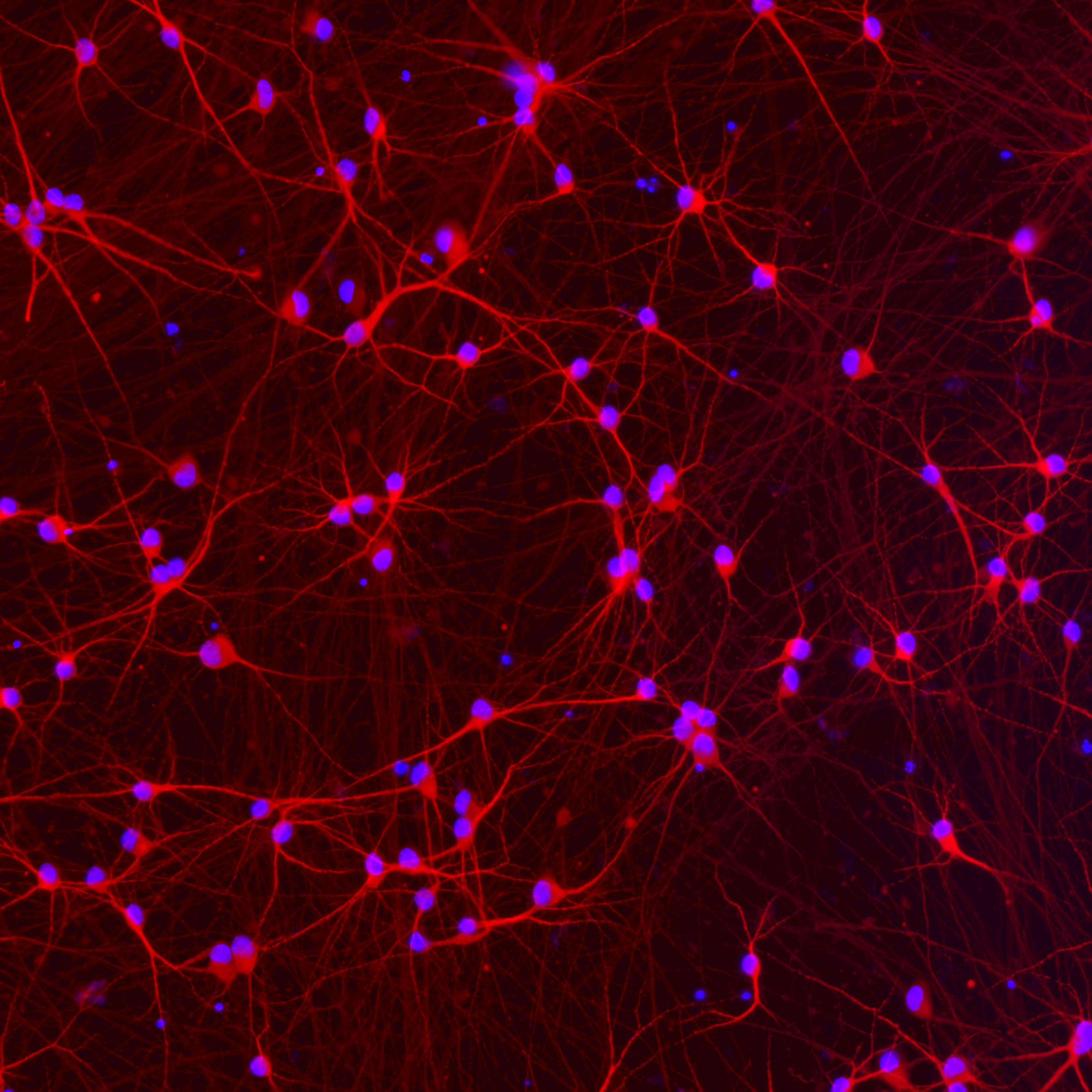
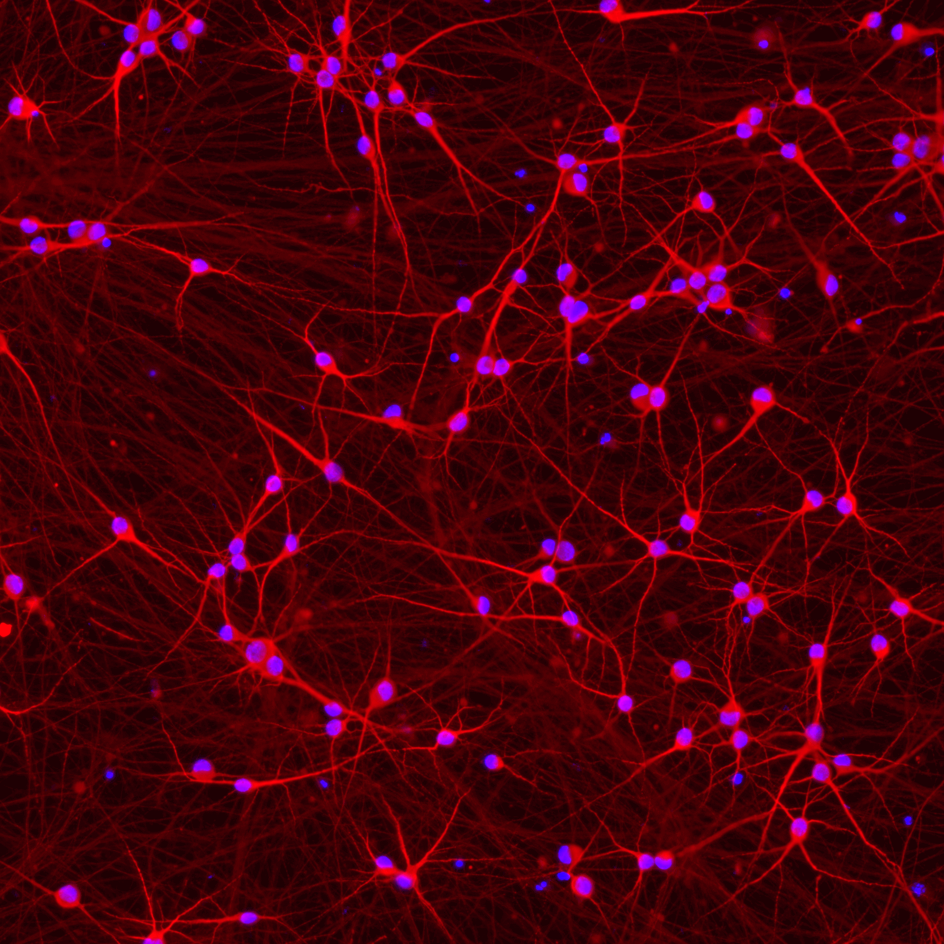
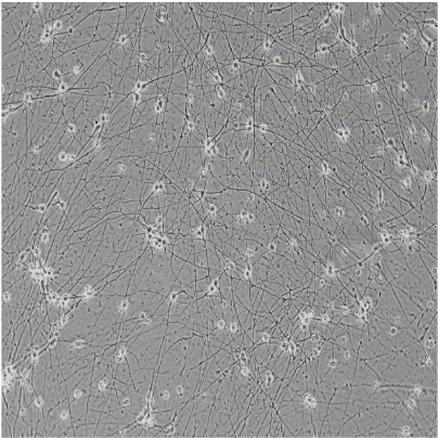
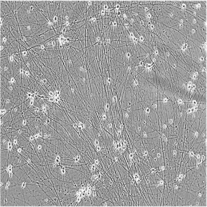
Human iPSC-derived glutamatergic neurons
-
Cryopreserved human iPSC-derived cells powered by opti-ox, that are ready for experiments in days
-
Ideal for studying excitatory signalling pathways, neurodegenerative diseases and neurotoxicology
-
Consistent, functional excitatory neurons, with no inhibitory neurons
Human iPSC-derived glutamatergic neurons
ioGlutamatergic Neurons display neuronal activity that matures over time
The function of ioGlutamatergic Neurons was investigated using the MaxTwo HD-MEA System.
The Axon Tracking Assay (left) shows examples of reconstructed axonal paths of travelling action potentials of individual iPSC-derived glutamatergic neurons. The assay reveals the spatial propagation of the neuronal action potential from the soma to distant axonal branches.
Total axon length (middle) and firing rate (right) increase over time, indicating that the cells are maturing. ioGlutamatergic Neurons were cultured with human iPSC-derived astrocytes.
Data courtesy of Charles River Laboratories and MaxWell Biosystems.
Rapid maturation of ioGlutamatergic Neurons leads to synchronised network activity by day 31
Raster plots generated using the MaxTwo HD-MEA System show the development of the neuronal network over time.
The plots show the dynamics of the network activity using 1,024 active electrodes. Each row represents an individual electrode and each blue dot indicates a spike detected at that electrode over a period of 300 seconds.
Spontaneous activity is observed at DIV 7. Clear synchronised bursting activity is observed by DIV 31, represented by blue vertical lines, followed by an overall drop in activity, seen as white lines.
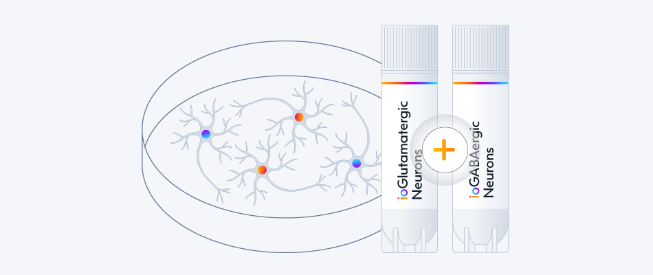
Download our poster to see additional data that shows how ioGABAergic Neurons form functional neuronal networks with ioGlutamatergic Neurons in the presence of astrocytes, and how the tri-culture responds to bicuculline and diazepam.
ioGlutamatergic Neurons offer a rapidly maturing functional system that can be used to assess neuronal networks and the impact of a drug treatment or intervention.
Data courtesy of Charles River Laboratories and MaxWell Biosystems.
ioGlutamatergic Neurons offer a robust, physiologically-relevant model for efficacy screening of candidate ASOs
Positive and negative control antisense oligonucleotides (ASOs) with gapmer chemistry were introduced into glutamatergic neurons by gymnosis. RT-qPCR was used to measure ASO-induced gene knockdown.
- Strong separation of the assay signal for positive control (blue) and negative control (orange) ASOs was observed for all plates tested (A).
- The positive control ASO induced ~90% knockdown of the target gene, shown by a decrease in the target gene expression (A) and higher Cp (or Ct) values for the target gene, indicating lower initial amount of the target sequence (B).
- There was no effect of the control ASOs on housekeeping gene expression as compared to vehicle-transfected controls (C).
- No marked intra- or inter-plate variability was observed between positive and negative control ASOs (A-C).
Data courtesy of Charles River Laboratories.
ioGlutamatergic Neurons show good suitability for high-throughput screening in 384-well format plates
Cytotoxicity CellTiter-Glo®️ (CTG) and TR-FRET (HTRF®️) assays for AKT serine/threonine kinase 1 (AKT) and Huntingtin (HTT) proteins were performed on ioGlutamatergic Neurons in 384-well plates treated with tool compound (cmp) at day 9 post-revival. Compound titration results in a concentration response curve for all three assays (mean±sd of 2 replicates). CTG assay on ioGlutamatergic Neurons shows an excellent average signal-to-background ratio and high suitability for HTS. HTRF assays on ioGlutamatergic Neurons show lower signals but with low variability, and could therefore also provide a suitable platform for HTS.
Data courtesy of Charles River Laboratories.
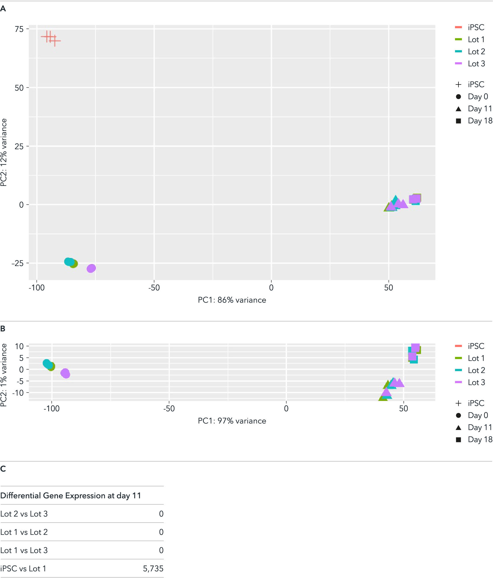
Whole transcriptome analysis demonstrates high lot-to-lot consistency across three manufactured lots of ioGlutamatergic Neurons
Bulk RNA-sequencing analysis was performed on three different lots of ioGlutamatergic Neurons on day 0, day 11 and day 18 post-revival. (A) A principal component analysis (PCA) to assess gene expression variance between three different manufactured lots showed a tight clustering of the samples at each timepoint, demonstrating high consistency between these lots. This lot-to-lot consistency of ioGlutamatergic Neurons will help reduce experimental variation and increase the reproducibility of experiments. (B) PCA without the parental non-induced hiPSC samples, highlighting the tight clustering of the day 11 as well as day 18 samples of the three different lots. (C) Differential expression test reveals no statistically significant differentially expressed (DE) genes across the three lots at day 11 (|logFC| > 0.5 and FDR < 0.01).
Colours represent the three lots of products; shapes represent the parental non-induced hiPSC line and different timepoints.
Expression levels for specific genes of interest can be requested by contacting our team at technical@bit.bio.
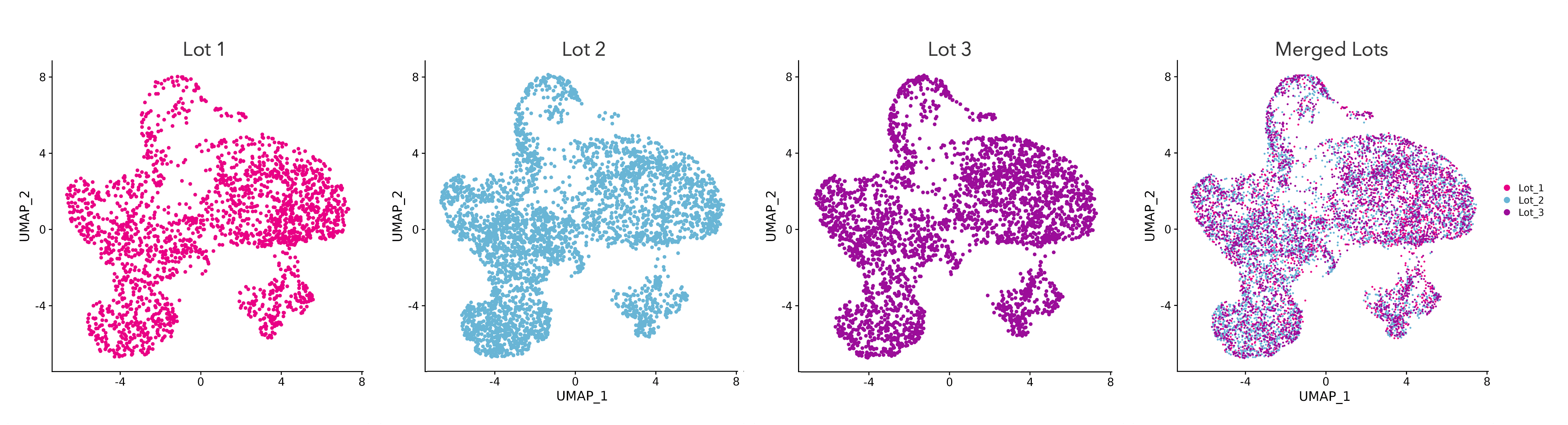
High lot-to-lot consistency is demonstrated by a consistent transcriptomic fingerprint across manufactured lots of ioGlutamatergic Neurons
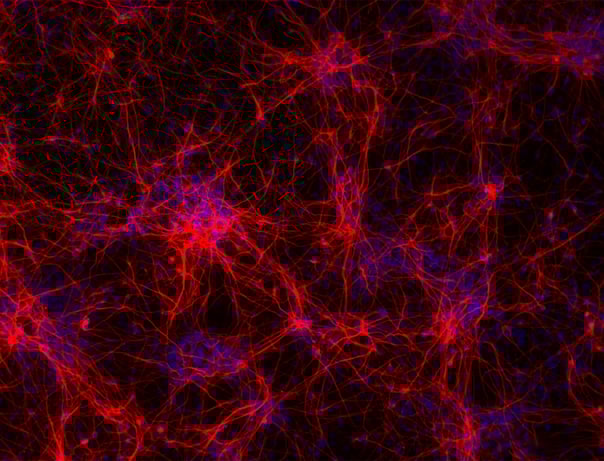
Single cell RNA-sequencing analysis was performed on three different lots of ioGlutamatergic Neurons on day 11. UMAP plots represent the cell-to-cell variation in gene expression profiles of cells, each dot representing an individual cell. Cells from each of the three lots are equally distributed across the body of the plot. Merging the UMAP plots creates a tight overlay, showing a strong transcriptional relationship between cells from three independently manufactured lots of ioGlutamatergic Neurons. Gene expression was assessed by 10x Genomics scRNA-sequencing.
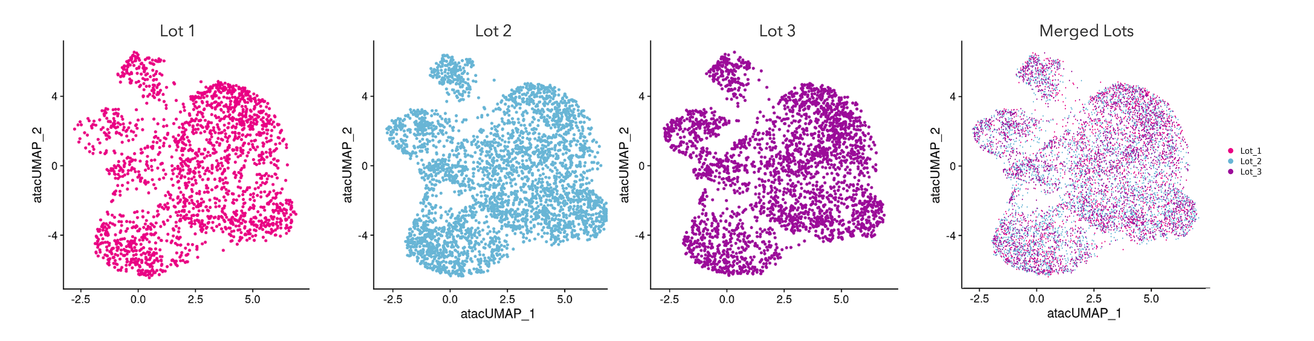
Single cell ATAC-sequencing shows a consistent transcriptomic fingerprint demonstrating high lot-to-lot consistency across manufactured lots of ioGlutamatergic Neurons
Single cell ATAC-sequencing analysis was performed on three different lots of ioGlutamatergic Neurons on day 11. Single cell ATAC-sequencing reveals regions of open chromatin to understand the gene regulatory landscape of individual cells. UMAP plots represent the cell-to-cell variation in chromatin accessibility of the cells, each dot representing a single cell. Cells from each of the three lots are equally distributed across the body of the plot. Merging the UMAP plots creates a tight overlay, showing a strong transcriptional relationship between cells from three independently manufactured lots of ioGlutamatergic Neurons. Gene expression was assessed by 10x Genomics scRNA-sequencing.
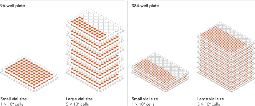
Vial limit exceeded
A maximum number of 20 vials applies. If you would like to order more than 20 vials, please contact us at orders@bit.bio.


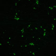
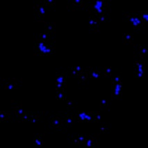
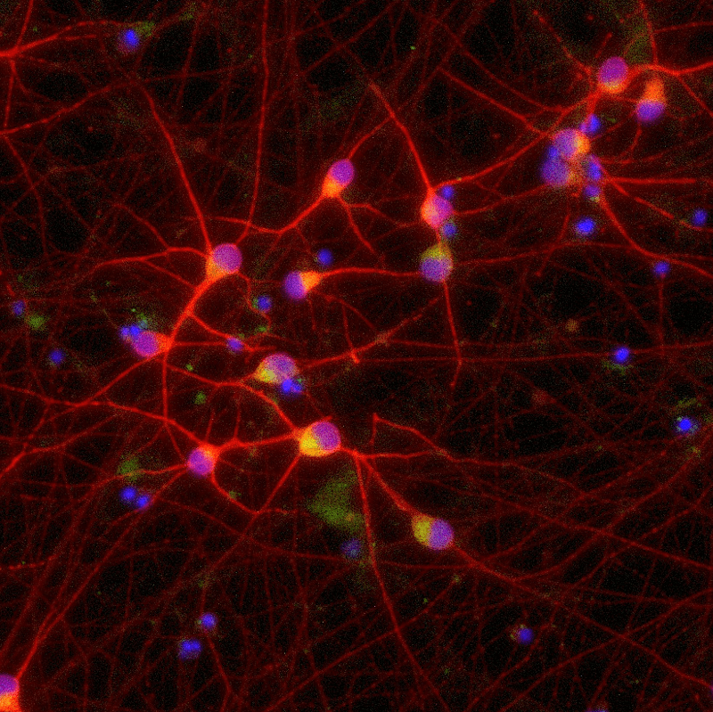
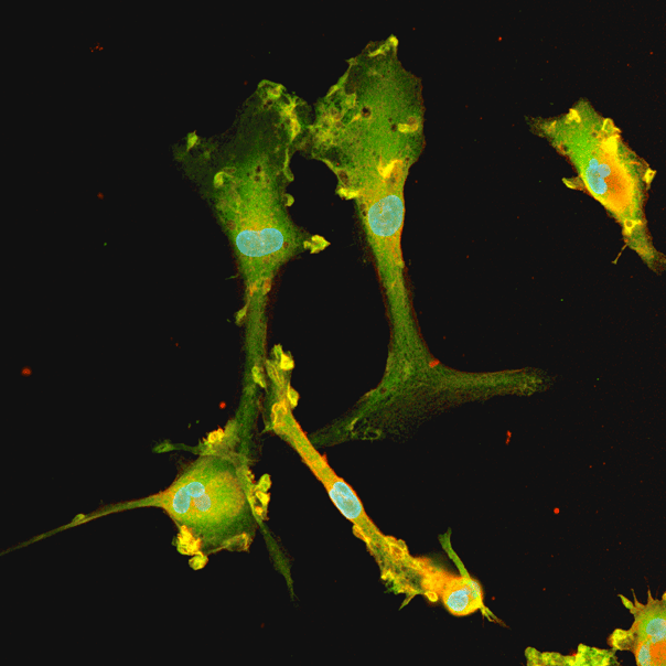
%20MAP2(r)%20DAPI(b).jpeg?width=213&height=213&name=bit-bio%20ioGlutamatergic%20Neurons%20-%20Day%2011%20-%20MERGE%20VGLUT2(g)%20MAP2(r)%20DAPI(b).jpeg)
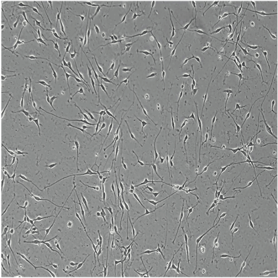
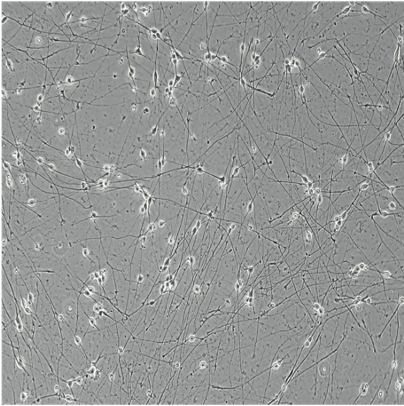


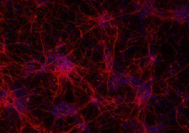


.png?width=1860&height=1260&name=bit.bio_3x2_ioGlutamatergic%20Neurons_MAP2_Hoescht_x20_hi.res%20(1).png)