19.09.2025 | Published by bit.bio
The human microglial clone 3 (HMC3) cell line has long been a staple for microglia research, widely used, readily available, and easy to culture. But as expectations around data relevance and human fidelity increase, many researchers are beginning to ask: Are HMC3 cells still the right model?
This blog walks through why labs are replacing HMC3 cells with ioMicroglia, a human iPSC-derived microglia developed by bit.bio. It compares the biology, highlights practical considerations, and shows how you can make the switch with minimal disruption.
What are HMC3 cells, and what are the limitations?
HMC3 is a human microglial cell line established in the 1990s through SV40 immortalisation. These cells have been widely adopted because they are easy to maintain and proliferate indefinitely. However, recent analyses reveal several critical issues1.
Known limitations of HMC3 cells
Limited microglial identity | HMC3 cells lack key microglia markers like P2RY12, TREM2, and IBA1. In fact, they express markers more typical of pericytes.
Reduced functionality | HMC3 cells demonstrate limited phagocytosis and a narrow, blunted cytokine response.
Phenotypic drift | As with many immortalised cell lines, long-term culture leads to inconsistency across labs and over passages.
If your goal is to model neuroinflammation, neuron-microglia interactions, or human-relevant microglial responses, the immortalised HMC3 cell line is a weak proxy.
ioMicroglia: Human-relevant phenotype, lot-to-lot consistency
ioMicroglia are derived from human iPSCs using opti-ox™ deterministic cell programming, resulting in:
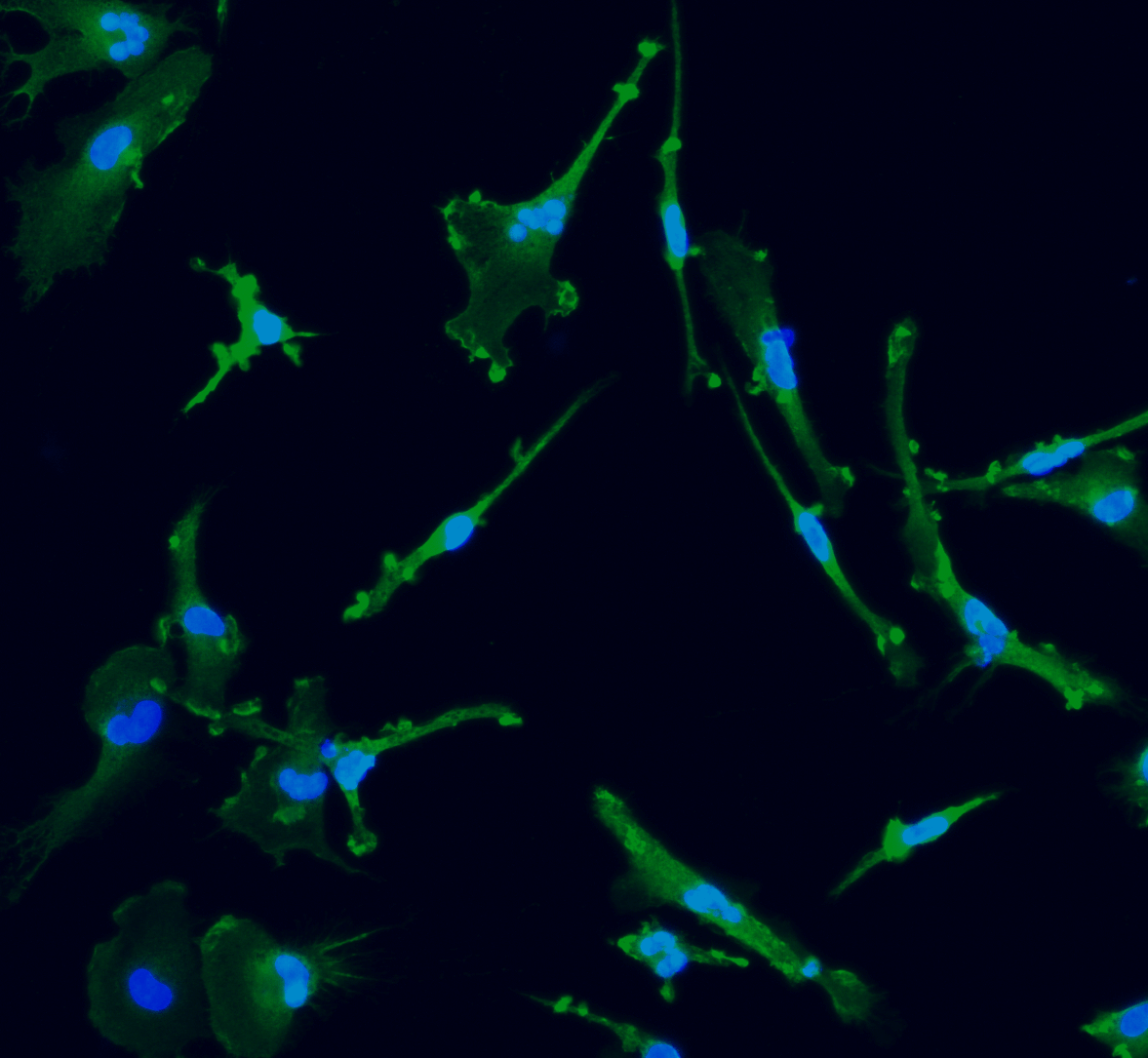
- >90% expression of canonical microglial markers like, P2RY12, CX3CR1, IBA1, and TREM2.
- Functional capabilities, including strong phagocytosis and broad cytokine secretion.
- Predictable, defined cultures without the risk of transformation or drift.
In short, they behave more like primary human microglia, while offering the consistency of a manufactured cell product.
|
Feature
|
HMC3 (immortalised)
|
ioMicroglia (iPSC-derived, deterministically programmed)
|
|
Identity
|
Pericyte-like, lacks key microglia markers |
|
|
Function
|
Low phagocytosis, weak cytokine release |
|
|
Proliferation
|
Divides indefinitely |
Post-mitotic after programming |
|
Reproducibility
|
Drift with passage number |
Consistent programming to a defined human microglia phenotype, lot-controlled, consistent functionality |
|
Co-culture
|
Medium compatibility, issues with neurons |
If you currently use HMC3 cells for phagocytosis or inflammatory readouts, and you are looking for a more phenotypically and physiologically relevant model, you can run the same assays with ioMicroglia, often with stronger and more reproducible results.
Do I have to change my workflows?
No. In most cases, you can apply your existing protocols with only minor adjustments:
- Phagocytosis assays: ioMicroglia engulf fluorescent beads or bacteria more efficiently than HMC3 cells. You may be able to reduce incubation time or cell numbers, making the assay more efficient.
- Microglial activation and cytokine release assays: ioMicroglia respond to LPS and amyloid-β with secretion of IL-6, IL-8, TNF-α, IL-1β, IL-12p70, and IL-10.
- Neuron-microglia co-culture: ioMicroglia can be co-cultured with human neurons using a hybrid medium. When cultured together, ioMicroglia develop ramified morphology and interact with neurons in a manner similar to what occurs in the CNS.
There is a short learning curve for handling and adopting workflows (e.g., gentle handling, day 10 differentiation). For an easy start, a video tutorial shows you step by step how to thaw, seed, and culture ioMicroglia.
In addition, we provide a detailed User Manual, Protocols, and Cell Culture Hacks that are designed for translational workflows and our Technical Support Team is always on hand.
Try ioMicroglia in your lab
See the difference in your own results, with our evaluation packs of ioMicroglia (3 vials for $995) so you can run side-by-side comparisons with your current HMC3 cell line setup. Each vial yields >1.0 million viable cells, enough for multiple assays.
The bottom line
If you are still using HMC3 cells, now is the time to re-evaluate. ioMicroglia offers a path to more physiologically relevant, functionally active, and consistent microglial models. With straightforward protocols and strong assay compatibility, the switch is easier than you might think.
Upgrading your microglial model is not about novelty; it is about getting better, translational data.
Reference
- Woolf, Z., Stevenson, T.J., Lee, K. et al. In vitro models of microglia: a comparative study. Sci Rep 15, 15621 (2025). https://doi.org/10.1038/s41598-025-99867-z